Purified anti-Alix Antibody

Pricing & Availability
Clone
3A9 (See other available formats)
Regulatory Status
RUO
Workshop
HCDM listed
Other Names
PDCD6IP, PDCD6 interacting protein, AIP1, Hp95
Isotype
Mouse IgG1, κ
Ave. Rating
Submit a Review
Product Citations
publications
3A9_Purified_Alix_Antibody_040419_updated.png
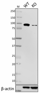
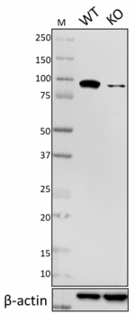
Total cell lysates (15 µg total protein) from HEK293T (WT) and HEK293T ALIX CRISPR/CAS9 knockout (KO) cells were resolved by 4-12% Bis-Tris gel electrophoresis, transferred to a nitrocellulose membrane, and probed with 2.0 µg/mL (1:250 dilution) of Purified anti-Alix Antibody, clone 3A9, overnight at 4°C. Proteins were visualized by chemiluminescence detection using HRP goat anti-mouse IgG Antibody (Cat. No. 405306) at a 1:3000 dilution. Direct-Blot™ HRP anti-β-Actin Antibody (Cat. No. 664804) was used as a loading control at a 1:25000 dilution (lower). Lane M: Molecular Weight marker.
  • 3A9_Purified_Alix_Antibody_040419_updated.png
    Total cell lysates (15 µg total protein) from HEK293T (WT) and HEK293T ALIX CRISPR/CAS9 knockout (KO) cells were resolved by 4-12% Bis-Tris gel electrophoresis, transferred to a nitrocellulose membrane, and probed with 2.0 µg/mL (1:250 dilution) of Purified anti-Alix Antibody, clone 3A9, overnight at 4°C. Proteins were visualized by chemiluminescence detection using HRP goat anti-mouse IgG Antibody (Cat. No. 405306) at a 1:3000 dilution. Direct-Blot™ HRP anti-β-Actin Antibody (Cat. No. 664804) was used as a loading control at a 1:25000 dilution (lower). Lane M: Molecular Weight marker.
  • 3A9_Purified_Alix_Antibody_2_031620
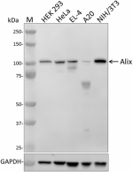
    Whole cell extracts (15 µg total protein) from the indicated cell lines were resolved by 4-12% Bis-Tris gel electrophoresis, transferred to a PVDF membrane, and probed with 1 µg/mL of purified anti-Alix antibody (clone 3A9) overnight at 4°C. Proteins were visualized by chemiluminescence detection using HRP goat anti-mouse IgG antibody (Cat. No. 405306) at a 1:3000 dilution. Direct-Blot™ HRP anti-GAPDH antibody (Cat. No. 607904) was used as a loading control at a 1:50000 dilution (lower). Lane M: Molecular weight marker.
Cat # Size Price Quantity Check Availability Save
634501 25 µg 90€
Check Availability


Need larger quantities of this item?
Request Bulk Quote
634502 100 µg 181€
Check Availability


Need larger quantities of this item?
Request Bulk Quote
Description

Alix (ALG-2-interacting protein X) is an adaptor protein that was first described for its capacity to bind to the calcium-binding protein ALG-2, the expression of which seemed necessary for cell death. The predicted molecular weight is approximately 96 kD. Alix contains an N-terminal Bro1 domain and a C-terminal proline-rich domain (PRD). The PRD contains multiple polyproline motifs, which are potential docking sites for proteins containing SH3 domains, interact with multiple cellular Alix-binding partners that are involved in apoptotic induction, endosomal sorting, and endocytosis. The Bro1 domain interacts with CHMP4b, which is also involved in endosomal sorting.

Product Details
Technical Data Sheet (pdf)

Product Details

Verified Reactivity
Human, Mouse
Reported Reactivity
Rat, Frog
Antibody Type
Monoclonal
Host Species
Mouse
Immunogen
Recombinant full-length human Alix protein
Formulation
This antibody is provided in phosphate-buffered solution, pH 7.2, containing 0.09% sodium azide.
Preparation
The antibody was purified by affinity chromatography.
Concentration
0.5 mg/ml
Storage & Handling
The antibody solution should be stored undiluted between 2°C and 8°C.
Application

WB - Quality tested
ICC, IP - Reported in the literature, not verified in house

Recommended Usage

Each lot of this antibody is quality control tested by Western blotting. Western blotting, suggested working dilution(s): Use 1 µg/ml antibody dilution buffer for each mini-gel. It is recommended that the reagent be titrated for optimal performance for each application.

Application Notes

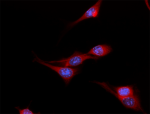
Clone 3A9 has been shown to be useful for Western blotting1, immunoprecipitation, and immunofluorescence2 staining of the human and mouse Alix protein.

Clone 3A9 shows strong specificity for mouse Alix.

Application References
  1. Morel L, et al. 2013. J Biol Chem. 288:7105. (WB) PubMed
  2. Pan S, et al. 2008. EMBO J. 27:2077. (IF)
  3. Ertunc ME, et al. 2015. J Lipid Res. 56:423. PubMed
Product Citations
  1. Yang C, et al. 2011. PLoS One. 6:e22517. PubMed
  2. Stempels FC, et al. 2023. J Cell Biol. 222:. PubMed
  3. Inouye KE, et al. 2023. JCI Insight. . PubMed
  4. Yim WW, et al. 2022. FEBS Lett. 596:991. PubMed
  5. Dong H, et al. 2022. Proc Natl Acad Sci U S A. 119:e2123247119. PubMed
  6. Tan JX, et al. 2022. Nature. 609:815. PubMed
  7. Jia X, et al. 2022. J Cell Biol. 221: . PubMed
  8. Burbidge K, et al. 2022. Autophagy. 18:1020. PubMed
  9. Suthar J, et al. 2020. Anal Chem. 92:4082. PubMed
  10. Jia J, et al. 2020. Dev Cell. 69:52. PubMed
  11. Yoon S, et al. 2017. Immunity. 47:51:00. PubMed
  12. Gulluni F, et al. 2021. Science. 374:eabk0410. PubMed
  13. Gorji-Bahri G, et al. 2021. Cell Biol Int. 45:1098. PubMed
  14. Morel L, et al. 2013. J Biol Chem. 288:7105. PubMed
  15. Valkov N, et al. 2021. Life Sci Alliance. 4:. PubMed
  16. Hüttenhain R et al. 2019. Cell Host Microbe. 26(1):86-99 . PubMed
  17. Moore D, et al. 2019. Stem Cells Int. 2019:8710180. PubMed
  18. Baba T, et al. 2021. Cell Death Dis. 12:322. PubMed
  19. Gärtner K, et al. 2018. Cancer Med. 11:39. PubMed
  20. Davis OB, et al. 2020. Developmental Cell. 56(3):260-276.e7. PubMed
  21. Ko J, et al. 2018. Lab Chip. 3.261805556. PubMed
  22. Skowyra ML, et al. 2018. Science. 360:6384. PubMed
  23. Gorji-Bahri G, et al. 2021. J Cell Biochem. 122:425. PubMed
  24. Andre-Gregoire G, et al. 2022. iScience. 25:105118. PubMed
  25. Gunasekaran M, et al. 2020. J Heart Lung Transplant. 39:379. PubMed
  26. Hiraga C, et al. 2021. Sci Rep. 11:3326. PubMed
  27. Chand S, et al. 2021. J Extracell Vesicles. 10:e12177. PubMed
  28. Claude-Taupin A, et al. 2021. Nat Cell Biol. 23:846. PubMed
  29. Ertunc M, et al. 2015. J Lipid Res. 56:423. PubMed
  30. Millarte V, et al. 2022. EMBO Rep. 23:e53429. PubMed
  31. Gunasekaran M, et al. 2018. J Immunol. 200:2535. PubMed
  32. Schiller LT, et al. 2018. Mol Ther Methods Clin Dev. 15:1630. PubMed
  33. Jia J, et al. 2020. Mol Cell. 951:77. PubMed
  34. Bæk R, et al. 2016. J Immunol Methods. 438:11-20. PubMed
RRID
AB_2162471 (BioLegend Cat. No. 634501)
AB_2162471 (BioLegend Cat. No. 634502)

Antigen Details

Structure
Belongs to a conserved family of proteins that have in common an N-terminal Bro1 domain and a C-terminal proline-rich domain. Predicted molecular weight approximately 96 kD.
Distribution

Cytoplasm. Alix is also abundant within extracellular vesicles known as exosomes, and enriched in virions that are released from cells under the direction of the HIV-1 Gag protein.

Function
Adaptor in signal transduction pathways. Involved in apoptosis and endosomal membrane trafficking and viral budding.
Interaction
ALG2, SH3GL2, H-Vps28, CHMP4B, CHMP4A, CHMP4C, SETA, CIN85, Src
Biology Area
Cell Biology
Antigen References

1. Wu Y, et al. 2000. Differentiation 67:139.
2. Missotten M, et al. 1999. Cell Death Differ. 6:124.
3. Odorizzi G. 2006. J Cell Sci. 119:3025.

Gene ID
10015 View all products for this Gene ID
UniProt
View information about Alix on UniProt.org

Related FAQs

There are no FAQs for this product.
Go To Top Version: 8    Revision Date: 09/19/2023

For Research Use Only. Not for diagnostic or therapeutic use.


This product is supplied subject to the terms and conditions, including the limited license, located at www.biolegend.com/terms) ("Terms") and may be used only as provided in the Terms. Without limiting the foregoing, BioLegend products may not be used for any Commercial Purpose as defined in the Terms, resold in any form, used in manufacturing, or reverse engineered, sequenced, or otherwise studied or used to learn its design or composition without express written approval of BioLegend. Regardless of the information given in this document, user is solely responsible for determining any license requirements necessary for user’s intended use and assumes all risk and liability arising from use of the product. BioLegend is not responsible for patent infringement or any other risks or liabilities whatsoever resulting from the use of its products.


BioLegend, the BioLegend logo, and all other trademarks are property of BioLegend, Inc. or their respective owners, and all rights are reserved.


8999 BioLegend Way, San Diego, CA 92121 www.biolegend.com
Toll-Free Phone: 1-877-Bio-Legend (246-5343) Phone: (858) 768-5800 Fax: (877) 455-9587

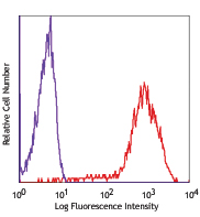
This data display is provided for general comparisons between formats.
Your actual data may vary due to variations in samples, target cells, instruments and their settings, staining conditions, and other factors.
If you need assistance with selecting the best format contact our expert technical support team.

ProductsHere

Login / Register
Remember me
Forgot your password? Reset password?
Create an Account