 Login / Register


- Clone
- W17025A (See other available formats)
- Regulatory Status
- RUO
- Other Names
- Histone Cluster 1, H2bb, H2B Histone Family-Member F, H2BFF, Histone H2B.1, H2B/F
- Isotype
- Rat IgG2a, λ
- Ave. Rating
- Submit a Review
- Product Citations
- publications

-

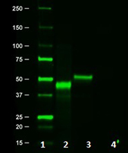
Total cell lysates (15 µg protein) from HeLa (lane 1), HaCaT (lane 2), and NIH/3T3 cells were resolved by 4-20% Tris-Glycine gel electrophoresis, transferred to nitrocellulose, and probed with the indicated concentration (µg/mL) of purified anti-Histone H2B (Clone W17025A) or a competitor’s clone used at the manufacturer’s recommended concentration (far right). Proteins were visualized by chemiluminescence detection using HRP goat anti-rat IgG (Cat. No. 405405) for clone W17025A and HRP goat anti-mouse IgG (Cat. No. 405306) for the competitor’s clone, each at a 1:3000 dilution. Direct-Blot™ HRP anti-β-actin Antibody (Cat. No. 643807) was used as a loading control at a 1:5000 dilution (lower). Lane M: Molecular weight ladder. -

Western blot of purified anti-Histone H2B antibody (clone W17025A). Lane 1: Molecular weight marker; Lane 2: 20 µg of Drosophila head lysate; Lane 3: 20 µg of Drosophila S2 (embryonic) cell lysate. The blot was incubated with 0.25 µg/mL of the primary antibody overnight at 4°C, followed by incubation with HRP-labeled goat anti-rat IgG (Cat. No. 405405). Enhanced chemiluminescence was used as the detection system. -

IHC staining of purified anti-Histone H2B antibody (clone W17025A) on formalin-fixed paraffin-embedded Drosophila brain tissue. Following antigen retrieval using Sodium Citrate H.I.E.R., the tissue was incubated with 1 µg/ml of the primary antibody overnight at 4°C, followed by incubation with Alexa Fluor® 594 goat anti- rat IgG (Cat No. 405422) for one hour at room temperature. Nuclei were counterstained with DAPI. A shows single channel image of purified anti-histone H2B antibody and B shows merged images with DAPI. The image was captured with a 40X objective. Scale bar: 50 µm
| Cat # | Size | Price | Quantity Check Availability | Save | ||
|---|---|---|---|---|---|---|
| 606301 | 25 µg | 81€ | ||||
| 606302 | 100 µg | 203€ | ||||
Histones are basic nuclear proteins that are responsible for the nucleosome structure of the chromosomal fiber in eukaryotes. Two molecules of each of the four core histones (H2A, H2B, H3, and H4) form an octamer, around which approximately 146 bp of DNA is wrapped in repeating units, called nucleosomes. Histones play central roles in transcription regulation, DNA repair, DNA replication and chromosomal stability. There are many variants of histones H2A and H2B. Specific H2A and H2B variants may preferentially associate with each other resulting in different combinations of variants and leading to the increased combinatorial complexity of nucleosomes. When being catalyzed by a series of histone modifying enzymes, histones may undergo various post-translational modifications such as acetylation, methylation, phosphorylation, ubiquitylation and SUMOylation. The dysregulation of histone modifying enzymes will alter the histone post-modification patterns and cause diverse diseases including cancers.
Product DetailsProduct Details
- Verified Reactivity
- Human, Mouse, Drosophila
- Antibody Type
- Monoclonal
- Host Species
- Rat
- Immunogen
- Human H2B1A peptide (111-127 a.a.)
- Formulation
- Phosphate-buffered solution, pH 7.2, containing 0.09% sodium azide.
- Preparation
- The antibody was purified by affinity chromatography.
- Concentration
- 0.5 mg/ml
- Storage & Handling
- The antibody solution should be stored undiluted between 2°C and 8°C.
- Application
-
WB - Quality tested
IHC-P - Verified - Recommended Usage
-
Each lot of this antibody is quality control tested by Western blotting. For Western blotting, the suggested use of this reagent is 0.25 - 2.0 µg per ml (1:250-2000). For immunohistochemistry on formalin-fixed paraffin-embedded tissue sections, a concentration range of 1.0 - 5.0 µg/ml is suggested. It is recommended that the reagent be titrated for optimal performance for each application.
- Application Notes
-
This clone is not recommended for ChIP (Chromatin Immunoprecipitation) assays (as determined by in-house testing).
Reactivity to Drosophila was only verified with the purified format. - Product Citations
-
- RRID
-
AB_2728464 (BioLegend Cat. No. 606301)
AB_2728465 (BioLegend Cat. No. 606302)
Antigen Details
- Structure
- 126 amino acids with a predicted molecular weight of approximately 14kD but observed at approximately 17kD as the highly positively charged Histone protein tends to migrate slower on SDS-PAGE gel.
- Distribution
-
Eukaryotic cells and nucleus.
- Function
- Histone H2B is one of the building blocks of nucleosome. It plays a central role in transcription regulation, DNA repair, DNA replication and chromosomal stability.
- Interaction
- Histone H2A
- Biology Area
- Cell Biology, Chromatin Remodeling/Epigenetics, DNA Repair/Replication
- Antigen References
-
1. Ma F, Zhang CY. 2016. Expert Rev. Mol. Diagn. 16:297.
2. Li J, et al. 2016. Biomed. Rep. 4:293.
3. Shaytan AK, et al. 2015. Curr. Opin. Struct. Biol. 32:48.
4. Wright DE, Kao CF. 2015. Epigenetics 10:122.
5. Kim J, et al. 2009. Cell. 137:459.
6. Cheung WL, et al. 2003. Cell. 113:507. - Gene ID
- 3018 View all products for this Gene ID
- UniProt
- View information about Histone on UniProt.org

Related FAQs
Other Formats
View All Histone Reagents Request Custom Conjugation| Description | Clone | Applications |
|---|---|---|
| Purified anti-Histone H2B | W17025A | WB,IHC-P |
Customers Also Purchased

Compare Data Across All Formats
This data display is provided for general comparisons between formats.
Your actual data may vary due to variations in samples, target cells, instruments and their settings, staining conditions, and other factors.
If you need assistance with selecting the best format contact our expert technical support team.


Follow Us