 Login / Register


- Clone
- W17155A (See other available formats)
- Regulatory Status
- RUO
- Other Names
- Lymphoid enhancer-binding factor 1, TCF1α, TCF10
- Isotype
- Rat IgG2b, κ
- Ave. Rating
- Submit a Review
- Product Citations
- publications
-

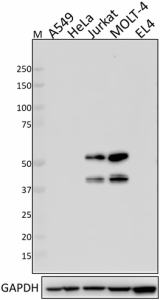
Total cell lysates (15 µg total protein) from A549 and HeLa cells (negative control), Jurkat, MOLT-4 and EL4 cells were resolved by 4-12% Bis-Tris gel electrophoresis, transferred to a PVDF membrane, and probed with 1.0 µg/mL (1:500 dilution) of Purified anti-LEF1 Antibody, clone W17155A, overnight at 4°C. Proteins were visualized by chemiluminescence detection using HRP Goat anti-rat IgG Antibody (Cat. No. 405405) at a 1:3000 dilution. Direct-Blot™ HRP anti-GAPDH Antibody (Cat. No. 607904) was used as a loading control at a 1:10000 dilution (lower). Lane M: Molecular Weight marker. Predicted LEF1 expression data was obtained from Human Protein Atlas. -
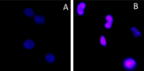
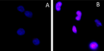
HeLa (negative control, panel A) and Jurkat (panel B) were fixed with 4% paraformaldehyde for 10 minutes, permeabilized with ice-cold methanol for 10 minutes, and blocked with 5% FBS for 60 minutes. Cells were then intracellularly stained with a 1:100 dilution (5 µg/mL) of Purified LEF1 Antibody, Clone W17155A overnight at 4°C, followed by incubation with Alexa Fluor® 594 goat anti-rat IgG (Cat. No. 405422) at 2.0 µg/mL. Nuclei were counterstained with DAPI and the image was captured with a 60X objective. -

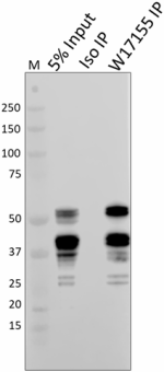
Whole cell extracts (300 µg total protein) prepared from Jurkat cells were immunoprecipitated overnight with 2.5 µg of Purified Rat IgG2b, κ Isotype Control Antibody (ISO IP, Cat. No. 400602) or Purified anti-LEF1 Antibody, clone W17155A (W17155A IP). The resulting IP fractions and whole cell extract input (5%) were resolved by 4-12% Bis-Tris gel electrophoresis, transferred to a PVDF membrane, and probed with a separate anti-LEF1 antibody raised with a different immunizing sequence. Lane M: Molecular Weight marker.
| Cat # | Size | Price | Quantity Check Availability | Save | ||
|---|---|---|---|---|---|---|
| 616001 | 25 µg | 81€ | ||||
| 616002 | 100 µg | 203€ | ||||
Lymphoid Enhancing Factor 1 (LEF1) is a transcription factor belonging to the TCF/LEF family. LEF1 was first identified as a protein binding to TCR-α enhancer and upregulates expression of T-cell antigen receptor α chain. LEF1 is involved in Wnt signaling pathways as a ubiquitous regulator. During Wnt signaling, translocation of β-catenin from the cytosol to the nucleus results in elevation of transcriptional activity of LEF1, which subsequently regulates multiple downstream genes.
Product DetailsProduct Details
- Verified Reactivity
- Human
- Antibody Type
- Monoclonal
- Host Species
- Rat
- Immunogen
- Partial recombinant human LEF1 protein corresponding to amino acid residues 116-283
- Formulation
- Phosphate-buffered solution, pH 7.2, containing 0.09% sodium azide.
- Preparation
- The antibody was purified by affinity chromatography.
- Concentration
- 0.5 mg/ml
- Storage & Handling
- The antibody solution should be stored undiluted between 2°C and 8°C.
- Application
-
WB - Quality tested
ICC, IP - Verified - Recommended Usage
-
Each lot of this antibody is quality control tested by Western blotting. For Western blotting, the suggested use of this reagent is 1.0 µg per ml. For immunocytochemistry, a concentration of 5.0 μg/ml is recommended. For immunoprecipitation, the suggested use of this reagent is 2.5 µg per ml. It is recommended that the reagent be titrated for optimal performance for each application.
- Application Notes
-
W17155A recognizes multiple isoforms of LEF1.
When tested for ICC, W17155A failed to stain PFA-fixed Jurkat cells permeabilized with methanol. We recommend Triton X-100 permeabilization. - RRID
-
AB_2814460 (BioLegend Cat. No. 616001)
AB_2814461 (BioLegend Cat. No. 616002)
Antigen Details
- Structure
- LEF1 is a 399 amino acid protein with a predicted molecular weight of 44 kD. Seven total isoforms have been reported, ranging in size from 23 to 44 kD.
- Distribution
-
Lymphoid tissue enriched/Nuclear localization
- Function
- Canonical Wnt signaling
- Biology Area
- Angiogenesis, Cell Biology, Immunology, Signal Transduction, Transcription Factors
- Molecular Family
- Nuclear Markers, TCRs
- Antigen References
-
1. Mallory MJ, et al. 2011. Mol. Cell. Biol. 31:2184.
2. Waterman ML, et al. 1991. Genes Dev. 5:656.
3. Hovanes K, et al. 2001. Nat Genet. 28:53.
4. Bruhn L, et al. 1997. Genes Dev. 11:640.
5. Nguyen A, et al. 2005. Int. J. Oncol. 27:949.
6. Petropoulos K, et al. 2008. J. Exp. Med. 205:515. - Gene ID
- 51176 View all products for this Gene ID
- UniProt
- View information about LEF1 on UniProt.org

Other Formats
View All LEF1 Reagents Request Custom Conjugation| Description | Clone | Applications |
|---|---|---|
| Purified anti-LEF1 | W17155A | WB,ICC,IP |
Compare Data Across All Formats
This data display is provided for general comparisons between formats.
Your actual data may vary due to variations in samples, target cells, instruments and their settings, staining conditions, and other factors.
If you need assistance with selecting the best format contact our expert technical support team.


Follow Us