- Clone
- W18298A (See other available formats)
- Regulatory Status
- RUO
- Other Names
- CFC, NS1, JMML, SHP2, BPTP3, PTP2C, METCDS, PTP-1D, SH-PTP2, SH-PTP3, Syp, Protein Tyrosine Phosphatase Non-Receptor Type 11, SH2 Domain-Containing Protein Tyrosine Phosphatase 2
- Isotype
- Rat IgG2a, λ
- Ave. Rating
- Submit a Review
- Product Citations
- publications

-

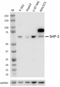
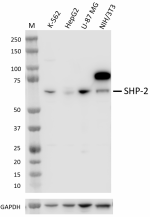
Whole cell extracts (15 µg total protein) from the indicated cell lines were resolved on a 4-12% Bis-Tris gel, transferred to a PVDF membrane, and probed with 1 µg/mL (1:500 dilution) of purified anti-SHP-2 (clone W18298A) overnight at 4°C. Proteins were visualized by chemiluminescence detection using HRP goat anti-rat IgG (Cat. No. 405405) at a 1:3000 dilution. Direct-Blot™ HRP anti-GAPDH (Cat. No. 607903) was used as a loading control at a 1:50000 dilution. Western-Ready™ ECL Substrate Plus Kit (Cat. No. 426317) was used as a detection agent. Lane M: Molecular weight marker -

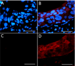
IHC staining of purified anti-SHP-2 (clone W18298A) on formalin-fixed paraffin-embedded human cerebellum tissue. Following antigen retrieval using 1X Tris-Buffered Saline with Tween-20 (final concentration 0.05M) (Cat. No. 925501), the tissue was incubated with blocking solution (negative control) (panels A and C) or 10 μg/mL of antibody (panels B and D) overnight at 4°C, followed by incubation with 2.5 μg/mL of Alexa Fluor® 647 goat anti-rat IgG (Cat. No. 405416) for one hour at room temperature. Nuclei were counterstained with DAPI (blue) (Cat. No. 422801) (panels A and B), and the slide was mounted with ProLong™ Gold Antifade Mountant. The image was captured with a 40X objective. Scale bar: 50 μm
| Cat # | Size | Price | Quantity Check Availability | Save | ||
|---|---|---|---|---|---|---|
| 609251 | 25 µg | 112€ | ||||
| 609252 | 100 µg | 295€ | ||||
Src homology-2-containing protein tyrosine phosphatase 2 (SHP-2) protein, encoded by the PTPN11 gene, is a well-known oncogenic protein tyrosine phosphatase. SHP-2 regulates cellular functions by dephosphorylating targets downstream of PD1, EGFRvIII, HGF receptor, and cytokine receptors. Cellular processes controlled by SHP-2 include mitotic cycle, cell growth, differentiation, and oncogenesis. SHP-2 has been recognized as a potential therapeutic target for cancers including breast cancer, lung cancer, leukemia, gastric cancer, and oral cancer.
Product DetailsProduct Details
- Verified Reactivity
- Human
- Antibody Type
- Monoclonal
- Host Species
- Rat
- Immunogen
- Recombinant fragment of human SHP-2
- Formulation
- Phosphate-buffered solution, pH 7.2, containing 0.09% sodium azide
- Preparation
- The antibody was purified by affinity chromatography.
- Concentration
- 0.5 mg/mL
- Storage & Handling
- The antibody solution should be stored undiluted between 2°C and 8°C.
- Application
-
WB - Quality tested
IHC-P - Verified - Recommended Usage
-
Each lot of this antibody is quality control tested by western blotting. For western blotting, the suggested use of this reagent is 0.25 - 1.0 µg/mL. For immunohistochemistry on formalin-fixed paraffin-embedded tissue sections, a concentration range of 5.0 - 10.0 µg/mL is suggested. It is recommended that the reagent be titrated for optimal performance for each application.
- Application Notes
-
This product cross reacts with mouse cell lines in Western-blot application but has poor selectivity. Clone A20016A is recommended for mouse reactivity.
This product is not recommended for immunocytochemistry. - RRID
-
AB_2927989 (BioLegend Cat. No. 609251)
AB_2927989 (BioLegend Cat. No. 609252)
Antigen Details
- Structure
- SHP-2 is a 593 amino acid protein with a molecular mass of 68 kD
- Distribution
-
Cytoplasm and Nucleus
- Function
- SHP-2 regulates cell proliferation, apoptosis, invasion, and metastasis
- Interaction
- SHP-2 interacts with PD1, EGFR, HGF receptor, RET, FGFR2, FGFR3, PI3K/AKT, and TLR3 signaling pathways.
- Cell Type
- Lymphocytes, T cells, Th1
- Biology Area
- Cell Biology, Cell Proliferation and Viability, Immuno-Oncology, Immunology
- Molecular Family
- Protein Kinases/Phosphatase
- Antigen References
-
- Hao C, et al. 2020. Oncogene. 39:7166-7180.
- Marie D, et al. 2008. Cell Signal. 20:453-9.
- Cheng Kui QU. 2000. Cell Research. 10:279-288.
- Gene ID
- 5781 View all products for this Gene ID
- UniProt
- View information about SHP-2 on UniProt.org
Other Formats
View All SHP-2 Reagents Request Custom Conjugation| Description | Clone | Applications |
|---|---|---|
| Purified anti-SHP-2 | W18298A | WB,IHC-P |
Compare Data Across All Formats
This data display is provided for general comparisons between formats.
Your actual data may vary due to variations in samples, target cells, instruments and their settings, staining conditions, and other factors.
If you need assistance with selecting the best format contact our expert technical support team.

 Login / Register


Follow Us