 Login / Register


- Clone
- A15158B (See other available formats)
- Regulatory Status
- RUO
- Other Names
- Signal transducer and activator of transcription 1 (STAT1), Transcription factor ISGF-3 components p91/p84
- Isotype
- Mouse IgG1, κ
- Ave. Rating
- Submit a Review
- Product Citations
- publications

-

Total lysates (15 µg protein) from untreated HeLa cells (lane 1) and HeLa cells treated with nocodazole (lane 2) were resolved by electrophoresis (4-12% Bis-Tris gel), transferred to nitrocellulose, and probed with 1:500 diluted (1 µg/mL) Purified anti-STAT1 Phospho (Ser727) Antibody, clone A15158B (upper) or 1:3000 diluted Purified anti-β-actin Antibody, clone Poly6221 (lower). Proteins were visualized by chemiluminescence detection using a 1:3000 diluted goat anti-mouse-IgG secondary antibody conjugated to HRP for the anti-STAT1 Phospho (Ser727) Antibody, and a donkey anti-rabbit IgG Antibody conjugated to HRP for anti-β-actin Antibody. -

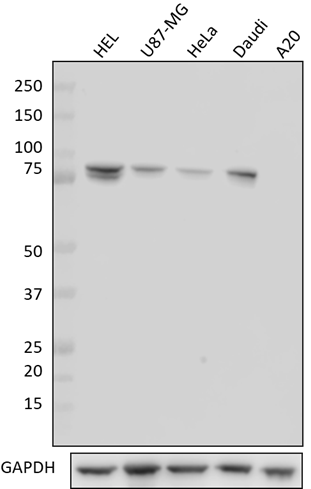
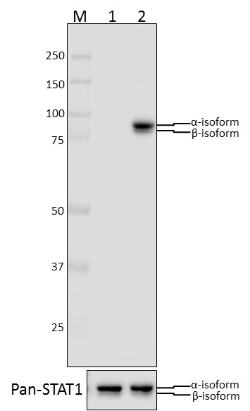
Total cell lysates (15 µg total protein) from NIH/3T3 cells untreated (-) or treated (+) with UV radiation (20 mJ for 5 seconds) were resolved by 4-12% Bis-Tris gel electrophoresis, transferred to a PVDF membrane, and probed with 1.0 µg/mL (1:500 dilution) of purified anti-STAT1 Phospho (Ser727) antibody (clone A15158B) overnight at 4°C. Proteins were visualized by chemiluminescence detection using HRP goat anti-mouse IgG antibody (Cat. No. 405306) at a 1:3000 dilution. Purified anti-STAT1 antibody (clone 10C4B40) (Cat. No. 661002), was used as a pan STAT1 loading control. Direct-Blot™ HRP anti-GAPDH antibody (Cat. No. 607904) was used as a loading control at a 1:25000 dilution (lower). Lane M: Molecular weight marker. -

HeLa cells were stimulated with (filled histogram) or without (open histogram) nocodozole for 24 hours, fixed with Fixation Buffer, permeabilized with True-Phos™ Perm Buffer, then intracellularly stained with purified anti-STAT1 Phospho (Ser727) antibody (clone A15158B), followed by anti- mouse IgG PE. -

Human peripheral blood lymphocytes were stimulated with (filled histogram) or without (open histogram) Cell Activation Cocktail (without Brefeldin A) for 15 minutes, fixed with Fixation Buffer, permeabilized with True-Phos™ Perm Buffer, then intracellularly stained with purified anti-STAT1 Phospho (Ser727) antibody (clone A15158B), followed by anti- mouse IgG PE. -

Two aliquots of HeLa cells in suspension, treated with (top) 200 ng/mL nocodazole for 24 hours or left un-treated (bottom) were adhered on poly-lysine pre-coated slides. Then they were fixed with 4% paraformaldehyde (PFA) for 15 minutes, permeabilized with 0.5% Triton X-100 for three minutes, and blocked with 5% FBS for 60 minutes. The cells were intracellularly stained with 2 µg/ml of purified anti-STAT1 Phospho (Ser727) antibody (clone A15158B) overnight at 4°C followed by Alexa Fluor® 594 (red) conjugated goat anti-mouse IgG for one hour at room temperature. Nuclei were counterstained with DAPI (blue). The image was captured with a 60X objective. -

Chromatin Immunoprecipitations (ChIP) were performed with cross-linked chromatin samples from 4 X 10⁶ of HeLa cells treated with Nocodazole with either A)1:50 dilution of Go-ChIP-Grade™ Purified anti-STAT1 Phospho (Ser727) Antibody (clone A15158B, Cat. No. 686413) or B) equal amount of Purified Mouse IgG1, κ Isotype Control Antibody (Clone MG1-45, Cat. No. 401401) by using Go-ChIP-Grade™ Protein G Enzymatic Kit (Cat. No. 699904). The enriched DNA was purified and quantified by real-time qPCR using primers targeting human IRF1 gene region or α-Satellite repeats. The amount of immunoprecipitated DNA in each sample is represented as signal relative to total amount of input chromatin.
| Cat # | Size | Price | Quantity Check Availability | Save | ||
|---|---|---|---|---|---|---|
| 686401 | 25 µg | 90€ | ||||
| 686402 | 100 µg | 193€ | ||||
STAT1, also known as signal transduction and activator of transcription 1, is a ubiquitously expressed cytoplasmic protein and is activated in response to cytokine signaling, including IFN-α, IFN-γ, EGF, PDGF, and IL-6. Upon activation, STAT1 is phosphorylated by receptor-associated kinases, translocates to the nucleus, and functions as a transcription factor. Two isoforms of STAT1, with apparent molecular weights of 88 and 91 kD, exist as a result of alternative RNA processing. STAT1 is involved in IFN-mediated immune responses, and STAT1-deficient mice are highly sensitive to bacterial and viral infections.
Product DetailsProduct Details
- Verified Reactivity
- Human, Mouse
- Antibody Type
- Monoclonal
- Host Species
- Mouse
- Immunogen
- Human STAT1 peptide phosphorylated at Ser 727.
- Formulation
- Phosphate-buffered solution, pH 7.2, containing 0.09% sodium azide.
- Preparation
- The antibody was purified by affinity chromatography.
- Concentration
- 0.5 mg/ml
- Storage & Handling
- The antibody solution should be stored undiluted between 2°C and 8°C.
- Application
-
WB - Quality tested
ICFC, ICC, ChIP - Verified - Recommended Usage
-
Each lot of this antibody is quality control tested by Western blotting. For Western blotting, the suggested use of this reagent is 0.5 - 2.5 µg/ml. For intracellular flow cytometry using our True-Phos™ Perm Buffer in Cell Suspensions Protocol, the suggested use of this reagent is ≤ 0.03 µg per million cells in 100 µl volume. For immunocytochemistry, a concentration range of 0.5 - 2.0 μg/ml is recommended. For ChIP applications, the suggested dilution is 1:50-1:100 by volume. It is recommended that the reagent be titrated for optimal performance for each application.
- Product Citations
-
- RRID
-
AB_2861070 (BioLegend Cat. No. 686401)
AB_2616883 (BioLegend Cat. No. 686402)
Antigen Details
- Structure
- 750 amino acids, predicted molecular weight of 87 kD; contains a SH2 domain responsible for homodimerization or heterodimerization.
- Distribution
-
Translocates to the nucleus when phosphorylated.
- Function
- Phosphorylated in response to cytokine signaling by receptor-associated kinases; translocates to the nucleus to act as a transcription factor. Mediates responses to type I (IFN-α/β) and II interferon (IFN-γ), EGF, PDGF, and IL-6.
- Interaction
- Forms a homodimer or heterodimers with other family members. Interacts with FAK, MCM3, MCM5, TRADD, BRCA1, KIT, IL-27R, IL-2Rβ, IL-2Rγ, IFNαβR, and c-Src.
- Biology Area
- Cell Biology, Signal Transduction
- Molecular Family
- Nuclear Markers, Phospho-Proteins
- Antigen References
-
1. Durbin JE, et al. 1996. Cell. 84:443.
2. Darnell JE Jr, et al. 1994. Science 264:1415.
3. Chen X, et al. 1998. Cell. 93:827.
4. Ramana CV, et al. 2000. Oncogene. 19:2619. - Gene ID
- 6772 View all products for this Gene ID
- UniProt
- View information about STAT1 Phospho Ser727 on UniProt.org
Related Protocols
- BioLegend’s Tools for Chromatin Immunoprecipitation (ChIP) Assays - Video
- Chromatin Immunoprecipitation (ChIP) Assay Protocol
- Immunocytochemistry Staining Protocol
- Western Blotting Protocol
- Intracellular Staining With True-Phos™ Perm Buffer in Cell Suspensions Protocol
- Intracellular Staining With True-Phos™ Perm Buffer in Whole Blood

Related FAQs
Other Formats
View All STAT1 Phospho (Ser727) Reagents Request Custom Conjugation| Description | Clone | Applications |
|---|---|---|
| Purified anti-STAT1 Phospho (Ser727) | A15158B | WB,ICFC,ICC,ChIP |
| PE anti-STAT1 Phospho (Ser727) | A15158B | ICFC |
| Alexa Fluor® 594 anti-STAT1 Phospho (Ser727) | A15158B | ICC |
| Alexa Fluor® 647 anti-STAT1 Phospho (Ser727) | A15158B | ICFC |
| Alexa Fluor® 488 anti-STAT1 Phospho (Ser727) | A15158B | ICFC |
| PE/Cyanine7 anti-STAT1 Phospho (Ser727) | A15158B | ICFC |
| PerCP/Cyanine5.5 anti-STAT1 Phospho (Ser727) | A15158B | ICFC |
Customers Also Purchased
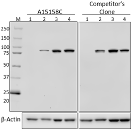
Compare Data Across All Formats
This data display is provided for general comparisons between formats.
Your actual data may vary due to variations in samples, target cells, instruments and their settings, staining conditions, and other factors.
If you need assistance with selecting the best format contact our expert technical support team.
-
Purified anti-STAT1 Phospho (Ser727)

Total lysates (15 µg protein) from untreated HeLa cells (lan... 
HeLa cells were stimulated with (filled histogram) or withou... 
Human peripheral blood lymphocytes were stimulated with (fil... 
Two aliquots of HeLa cells in suspension, treated with (top)... 
Total cell lysates (15 µg total protein) from NIH/3T3 cells ... 
Chromatin Immunoprecipitations (ChIP) were performed with cr... -
PE anti-STAT1 Phospho (Ser727)

HeLa cells were stimulated with (filled histogram) or withou... 
Human peripheral blood lymphocytes were stimulated with (fil... 
NIH/3T3 cells were treated with (filled histogram) or withou... -
Alexa Fluor® 594 anti-STAT1 Phospho (Ser727)

Two aliquots of HeLa cells in suspension, treated with 200 n... -
Alexa Fluor® 647 anti-STAT1 Phospho (Ser727)

HeLa cells were stimulated with (filled histogram) or withou... 
Human peripheral blood lymphocytes were stimulated with (fil... -
Alexa Fluor® 488 anti-STAT1 Phospho (Ser727)

HeLa cells were stimulated with (filled histogram) or withou... 
Human peripheral blood lymphocytes were stimulated with (fil... -
PE/Cyanine7 anti-STAT1 Phospho (Ser727)

HeLa cells were stimulated with (filled histogram) or withou... 
Human peripheral blood lymphocytes were stimulated with (fil... -
PerCP/Cyanine5.5 anti-STAT1 Phospho (Ser727)

HeLa cells were treated with (filled histogram) or without (... 
Human peripheral blood lymphocytes were treated with (filled...


Follow Us