Purified anti-Tubulin β-3 (TUBB3) Antibody (Previously Covance catalog# PRB-435P)

Pricing & Availability
Clone
Poly18020 (See other available formats)
Regulatory Status
RUO
Other Names
CDCBM, CDCBM1, CFEOM3, CFEOM3A, FEOM3, TUBB4, Tubulin beta-3 chain, tubulin beta-III, TUBB3, tubulin beta-4 chain, class III beta-tubulin
Previously
Covance Catalog# PRB-435P
Isotype
Rabbit Polyclonal IgG
Ave. Rating
Submit a Review
Product Citations
publications
A_Poly18020_PURE_Tubulin_beta3_IHCP_1_032918
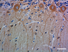
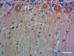
IHC staining of purified anti-Tubulin β-3 (TUBB3) antibody (Poly18020) on formalin-fixed paraffin-embedded rat cerebellum. Following antigen retrieval using Sodium Citrate H.I.E.R., the tissue was incubated with 0.1 µg/mL of the primary antibody overnight at 4°C. BioLegend’s Ultra Streptavidin (USA) HRP Detection Kit (Multi-Species, DAB, Cat. No. 929901) was used for detection followed by hematoxylin counterstaining, according to the protocol provided. The image was captured with a 40X objective. Scale bar: 50 µm
  • A_Poly18020_PURE_Tubulin_beta3_IHCP_1_032918
    IHC staining of purified anti-Tubulin β-3 (TUBB3) antibody (Poly18020) on formalin-fixed paraffin-embedded rat cerebellum. Following antigen retrieval using Sodium Citrate H.I.E.R., the tissue was incubated with 0.1 µg/mL of the primary antibody overnight at 4°C. BioLegend’s Ultra Streptavidin (USA) HRP Detection Kit (Multi-Species, DAB, Cat. No. 929901) was used for detection followed by hematoxylin counterstaining, according to the protocol provided. The image was captured with a 40X objective. Scale bar: 50 µm
  • B_Poly18020_PURE_Tubulin_beta3_IHCP_2_032918
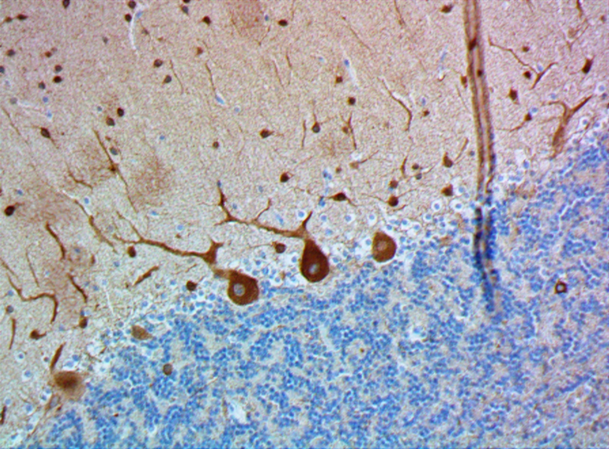
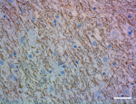
    IHC staining of purified anti-Tubulin β-3 (TUBB3) antibody (Poly18020) on formalin-fixed paraffin-embedded human cerebellum. Following antigen retrieval using Sodium Citrate H.I.E.R., the tissue was incubated with 0.1 µg/mL of the primary antibody overnight at 4°C. BioLegend’s Ultra Streptavidin (USA) HRP Detection Kit (Multi-Species, DAB, Cat. No. 929901) was used for detection followed by hematoxylin counterstaining, according to the protocol provided. The image was captured with a 40X objective. Scale bar: 50 µm
  • C_Poly18020_PURE_Tubulin_beta3_IHCP_3_032918
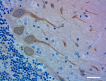
    IHC staining of purified anti-Tubulin β-3 (TUBB3) antibody (Poly18020) on formalin-fixed paraffin-embedded human cerebellum. Following antigen retrieval using Sodium Citrate H.I.E.R., the tissue was incubated with 0.1 µg/mL of the primary antibody overnight at 4°C. BioLegend’s Ultra Streptavidin (USA) HRP Detection Kit (Multi-Species, DAB, Cat. No. 929901) was used for detection followed by hematoxylin counterstaining, according to the protocol provided. The image was captured with a 40X objective. Scale bar: 50 µm
  • Poly18020_PURE_Tubulin_beta3_WB_021915.jpg
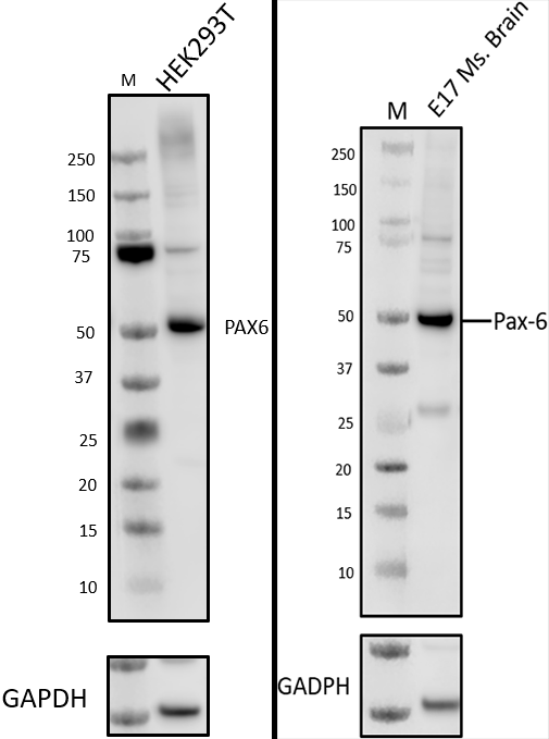
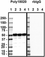
    Western blot of purified anti-Tubulin β-3 (TUBB3) antibody (Poly18020). Lane 1: Molecular weight marker; Lane 2: 20 µg of human brain lysate; Lane 3: 20 µg of mouse brain lysate; Lane 4: 20 µg of rat brain lysate. The blots were incubated with 0.1 µg/mL of Poly18020 or Rabbit IgG overnight at 4°C, followed by incubation with HRP-labeled anti-rabbit IgG (Cat. No. 410406). Enhanced chemiluminescence was used as the detection system.
Cat # Size Price Save
802001 100 µL ¥95,260
Description

Tubulin is the main component of microtubules. In adults, tubulin beta 3 (TUBB3) is primarily expressed in neurons and is commonly used as a neuronal marker. It plays an important role in neuronal cell proliferation and differentiation. Mutations in this gene cause congenital fibrosis of the type 3 extraocular muscles. Tubulin beta 3 (TUBB3) is also found in a wide range of tumors. Studies indicate that it is a predictive and prognostic marker in various tumors.

Product Details
Technical data sheet

Product Details

Verified Reactivity
Human, Mouse, Rat
Antibody Type
Polyclonal
Host Species
Rabbit
Immunogen
This antibody was raised against microtubules derived from rat brain.
Formulation
Phosphate-buffered solution + 0.03% Thimerosal + 50% Glycerol.
Preparation
The antibody was purified by affinity chromatography.
Concentration
1.0 mg/ml
Storage & Handling
Store between 2°C and 8°C.
Application

IHC-P - Quality tested
WB - Verified
ICC - Reported in the literature, not verified in house

Recommended Usage

Each lot of this antibody is quality control tested by formalin-fixed paraffin-embedded immunohistochemical staining. For immunohistochemistry, a concentration range of 0.1 - 0.5 µg/mL is suggested. For Western blotting, the suggested use of this reagent is 0.1 - 0.5 µg/mL. It is recommended that the reagent be titrated for optimal performance for each application.

Application Notes

Additional reported applications (for the relevant formats) include: immunocytochemistry (ICC).

This antibody is well characterized and highly reactive to neuron specific Class III ß-Tubulin (ßIII), but does not identify ß-tubulin found in glial cells. This product may contain other non-IgG subtypes.

Application References

(PubMed link indicates BioLegend citation)
  1. Feuer R, et al. 2005. J. Neurosci. 25(9):2434-44.
  2. Mozzetti S, et al. 2005. Clin. Cancer Res. 11(1):298-305.
  3. Feuer R, et al. 2003. Am. J. Pathol. 163(4):1379-93.
  4. Zhang XM, Yang XJ. 2001. Development. 128(6):943-57.
  5. Ma M, et al. 2012. Neurobiol Dis. 56:34. (WB) PubMed
  6. Tseung G, et al. 2011. J. Virol. 85:5718. (IF) PubMed
  7. Shimada IS, et al. 2012. J Neurosci. 32:7926. (IHC) PubMed
  8. Flores-Otero J, et al. 2007. J. Neurosci. 27:14023. (IF) PubMed
Product Citations
  1. Tseng K, et al. 2017. eNeuro. 10.1523/ENEURO.0214-17.2017. PubMed
  2. Traxler L, et al. 2022. Cell Metab. 34:1248. PubMed
  3. Asamitsu S, et al. 2023. Sci Adv. 9:eade2035. PubMed
  4. Weng ZK, et al. 2023. Biomolecules. 13:. PubMed
  5. Tian JL, et al. 2023. Cells. 12:. PubMed
  6. Drobny S, et al. 2023. Transl Neurodegener. 12:31. PubMed
  7. Chow A, Morshead C 2016. Stem Cell Res. 16:79-87. PubMed
  8. Son M, et al. 2016. Exp Mol Med. 48: 232. PubMed
  9. Hoshino N, et al. 2021. Journal of Comparative Neurology. 529:3633. PubMed
  10. Ahangari N, et al. 2021. J Neuropathol Exp Neurol. 80:944. PubMed
  11. Samarasinghe RA, et al. 2021. Nat Neurosci. 24:1488. PubMed
  12. Yates PL, et al. 2021. Cell Mol Life Sci. 78:6941. PubMed
  13. Hodgetts SI, et al. 2022. Neural Regen Res. 17:1376. PubMed
  14. Muralidharan H, et al. 2022. J Neurosci. 42:2149. PubMed
  15. Jabali A, et al. 2022. EMBO Rep. 23:e54027. PubMed
  16. Reis LTC, et al. 2022. Environ Toxicol. 37:2044. PubMed
  17. Rodriguez CM, et al. 2022. Cell Rep. 41:111505. PubMed
  18. Matsubayashi J, et al. 2023. J Neurochem. 164:29. PubMed
  19. Libanori A, et al. 2023. Adv Mater. 35:e2206933. PubMed
  20. Shimizu K, et al. 2022. Cells. 11: . PubMed
  21. Olmsted ZT, et al. 2022. STAR Protoc. 3:101898. PubMed
  22. Mogas Barcons A, et al. 2022. Materials (Basel). 16: . PubMed
  23. Yates PL, et al. 2023. Front Cell Neurosci. 16:979652. PubMed
  24. Genchi A, et al. 2023. Nat Med. 29:75. PubMed
  25. Chiu YJ, et al. 2023. Biomol Ther (Seoul). :. PubMed
  26. Willumsen N, et al. 2023. Brain Commun. 5:fcac321. PubMed
  27. Yang OJ, et al. 2023. Front Neurosci. 17:1001007. PubMed
  28. Stojkovska I, et al. 2022. Neuron. 110:436. PubMed
  29. Prieto Huarcaya S, et al. 2022. Autophagy. 18:1127. PubMed
  30. Arber C, et al. 2019. Brain Commun. 1:fcz024. PubMed
  31. Jiang Y, et al. 2018. J Biol Chem. 293:19317. PubMed
  32. Stokowska A, et al. 2017. Brain. 140:353. PubMed
  33. Kim JS, et al. 2020. Immunity. 54(1):176-190.e7. PubMed
  34. Tian E, et al. 2016. Cell Rep. 16: 781-792. PubMed
  35. Baleriola J, et al. 2016. Sci Rep. 6: 25928. PubMed
  36. Shimada I, et al. 2012. J Neurosci. 32:7926-7940. PubMed
  37. Eltahir MK, et al. 2022. PLoS One. 17:e0269683. PubMed
  38. Pardieck J, et al. 2021. Stem Cells Dev. 30:816. PubMed
  39. Kim S, et al. 2021. Nat Commun. 12:1807. PubMed
  40. Brosig A, et al. 2019. Cell Rep. 29:2028. PubMed
  41. Girouard MP, et al. 2020. eneuro. 7(2):ENEURO. PubMed
  42. Krishnan V, et al. 2016. J Neuropathol Exp Neurol. 75: 464 - 478. PubMed
  43. Meng QT, et al. 2021. Elife. 10:. PubMed
  44. Olmsted ZT, et al. 2022. Front Cell Neurosci. 15:725195. PubMed
  45. Binder J, et al. 2022. Commun Biol. 5:125. PubMed
  46. Nichterwitz S, et al. 2020. Genome Res. 30:1083. PubMed
  47. Vogt M, et al. 2021. Theranostics. 11:5569. PubMed
  48. Pires G, et al. 2021. Brain Commun. 3:fcab021. PubMed
  49. Xie Y, et al. 2018. Stem Cell Res. 9:2010. PubMed
  50. Uezono N, et al. 2018. Stem Cells. 36:737. PubMed
  51. Tang SC, et al. 2017. Diabetologia. 61:158. PubMed
  52. MOSES C, et al. 2020. Sci Rep. 10:11393. PubMed
  53. Nayak G, et al. 2020. Cell Reports. 30(3):672-686.e8.. PubMed
  54. Brykczynska U, et al. 2016. Stem Cell Reports. 7:1059-1071. PubMed
  55. Omichi R, et al. 2020. Mol Ther Methods Clin Dev. 1.51875. PubMed
  56. Hastings RL, et al. 2020. Sci Rep. 10:11132. PubMed
  57. Hoffman LF, et al. 2018. J Assoc Res Otolaryngol. 19:33. PubMed
  58. Carlin D et al. 2018. eNeuro. 5(2) pii: ENEURO. PubMed
  59. Jo S, et al. 2021. Stem Cell Research. 56:102524. PubMed
  60. Gong C, et al. 2021. Cell Reports. 34(12):108889. PubMed
  61. Kurihara Y, et al. 2020. Journal of Neurochemistry. 155(3):285-299. PubMed
  62. Dvoriantchikova G, et al. 2022. Sci Rep. 12:17152. PubMed
  63. Sakaguchi H, et al. 2019. Stem Cell Reports. 13:458. PubMed
  64. Voronova A et al. 2017. Neuron. 94(3):500-516 . PubMed
  65. Sabelström H, et al. 2019. Cell Rep. 28:2064. PubMed
  66. Boland M, et al. 2017. Brain. 10.1093/brain/aww357. PubMed
  67. Ou MY, et al. 2021. Cell Reports. 35(13):109290. PubMed
  68. Mertens J, et al. 2021. Cell Stem Cell. 28:1533. PubMed
  69. Verstraelen P, et al. 2017. Front Cell Neurosci. 11:173. PubMed
  70. Kim H et al. 2017. Stem cell research. 26:72-75 . PubMed
  71. Li H, et al. 2019. iScience. 19:559. PubMed
  72. Otani T, et al. 2016. Cell Stem Cell. 18: 467-480. PubMed
  73. van Niekerk EA, et al. 2022. Cell Rep Methods. 2:100255. PubMed
  74. Avraham O, et al. 2021. Elife. 10:. PubMed
  75. Simone R, et al. 2021. Nature. 594:117. PubMed
  76. Lasagni Vitar RM, et al. 2021. Invest Ophthalmol Vis Sci. 62:26. PubMed
  77. Reddy A, et al. 2020. Cell. 183:62. PubMed
  78. Benoy V, et al. 2018. Brain. 10.1093/brain/awx375. PubMed
  79. Chabrat A, et al. 2019. Stem Cells Int. 2019:2945435. PubMed
  80. Yang C, et al. 2020. Neuron. 105:276. PubMed
  81. Suter TACS, et al. 2020. Cell Rep. 30:1164. PubMed
  82. Macpherson P, et al. 2015. Development. 142: 4038 - 4048. PubMed
  83. Ferguson M, et al. 2013. Neurobiol Dis. 56:34-46. PubMed
  84. Yau K, et al. 2016. J Neurosci. 36: 1071 - 1085. PubMed
  85. Cui Q, et al. 2016. Nat Commun. 7:10637. PubMed
  86. Bosze B, et al. 2020. J Neurosci. 40:1501. PubMed
  87. Iannielli A, et al. 2019. Cell Rep. 29:4646. PubMed
  88. Bowles KR, et al. 2019. PLoS One. 14:e0213374. PubMed
  89. Toda T, et al. 2017. Cell Stem Cell. 21:618. PubMed
  90. Adams MT, et al. 2021. eLife. 0.416666666666667. PubMed
  91. Qiang L et al. 2018. Current biology : CB. 28(13):2181-2189 . PubMed
  92. Meijer M et al. 2019. Cell Rep. 27(7):2199-2211 . PubMed
  93. Rhee HJ et al. 2019. Cell Rep. 27(7):2212-2228 . PubMed
  94. Makar T, et al. 2018. Stem Cell Res Ther. 9:127. PubMed
  95. Zahr SK et al. 2018. Neuron. 97(3):520-537 . PubMed
  96. Amador A, et al. 2020. Brain. 143:2039. PubMed
  97. Sugiyama T, et al. 2021. iScience. 24(7):102758. PubMed
  98. Bhaskaran S, et al. 2020. Aging Cell. 19:e13225. PubMed
  99. Finno CJ, et al. 2022. Front Cell Neurosci. 16:853035. PubMed
  100. Newland B, et al. 2021. ACS Chem Neurosci. 12:1178. PubMed
  101. Cora V, et al. 2019. Cells. 8:5. PubMed
  102. Grove M, et al. 2020. eLife. 9:e50138.. PubMed
  103. Alvarez–Lindo N, et al. 2019. Sci Rep. 9:18486. PubMed
  104. Tiwari D et al. 2019. Neurobiol Dis. 1871:48:00 . PubMed
  105. Carlin D et al. 2019. eNeuro. 6(3) pii: ENEURO. PubMed
  106. Li H, et al. 2018. Hum Mutat. 39:550. PubMed
  107. Huang S, et al. 2020. Cell. 184(2):441-459.e25. PubMed
  108. Lin P, et al. 2016. Am J Physiol Gastrointest Liver Physiol. 311: G412 - G422. PubMed
  109. Tsueng G, et al. 2011. J Virol. 85:5718-5732. PubMed
  110. Francistiová L, et al. 2022. Front Mol Neurosci. 14:793769. PubMed
  111. Iennaco R, et al. 2022. Cell Death Differ. 29:293. PubMed
  112. Bjorke B, et al. 2021. Dev Biol. 476:272. PubMed
  113. Ou J et al. 2018. Cell. 173(4):851-863 . PubMed
  114. Hsu J, et al. 2019. Nat Commun. 10:2939. PubMed
  115. Chanda S et al. 2019. Cell Stem Cell. 25(1):103-119 . PubMed
  116. Peitz M, et al. 2020. STAR Protoc. 1:100038. PubMed
  117. Watanabe S, et al. 2016. EMBO Mol Med. 8(12):1421-1437. PubMed
  118. Cates K, et al. 2020. Cell Stem Cell. 28(1):127-140.e9. PubMed
  119. Smith LA, et al. 2021. Front Cell Dev Biol. 9:667246. PubMed
  120. Mao Y, et al. 2021. Cell Prolif. 54:e13090. PubMed
  121. Olmsted ZT, et al. 2022. iScience. 25:104486. PubMed
  122. Vahsen BF, et al. 2022. Sci Rep. 12:12606. PubMed
  123. Barry DM, et al. 2020. Nat Commun. 11:1397. PubMed
  124. Hattori Y, et al. 2020. Nat Commun. 11:1631. PubMed
  125. Duncan DS, et al. 2018. Front Neurosci. 0.9875. PubMed
  126. Martínez JC et al. 2019. Neuron. 104(5):931-946 . PubMed
  127. Mickle AD, et al. 2019. Nature. 565:361. PubMed
  128. Kawakami Y et al. 2018. The Journal of Neuroscience. 38(10):2589-2604 . PubMed
  129. Vicidomini C, et al. 2016. Mol Psychiatry. 10.1038/mp.2016.30. PubMed
  130. Flitsch LJ, et al. 2022. Sci Rep. 12:8356. PubMed
  131. Pahl MC, et al. 2021. Nat Commun. 12:6749. PubMed
  132. Kim H, et al. 2020. Sci Signal. 13: . PubMed
  133. Shah K, et al. 2019. J Mol Cell Biol. N/A. PubMed
  134. Sar Shalom H, et al. 2019. Elife. 8:e41162. PubMed
  135. Facchin C, et al. 2020. Theranostics. 10:3518. PubMed
  136. Flores-Otero J, et al. 2007. J Neurosci. 27:14023-14034. PubMed
  137. Zhang L, et al. 2016. Neuroscience. 337:88-97. PubMed
  138. Arber C, et al. 2021. Cell Reports. 34(2):108615. PubMed
  139. Shang Y, et al. 2018. J Neurosci. 38:4006. PubMed
  140. Jean-Louis T, et al. 2018. Neurobiol Aging. 62:130. PubMed
  141. Besusso D, et al. 2020. Stem Cell Res. 102016:49. PubMed
  142. Baker L, et al. 2021. JCI Insight. 6:. PubMed
  143. Hung COY et al. 2018. Cell reports. 25(13):3647-3660 . PubMed
  144. Jeon H, et al. 2018. J Clin Med. 7:. PubMed
  145. Arber C, et al. 2020. Mol Psychiatry. 25:2919. PubMed
  146. Smith-Anttila CJA, et al. 2021. Developmental Biology. 476:18-32. PubMed
  147. Hung C, et al. 2021. Cell Reports. 35(11):109259. PubMed
  148. Ornaghi F, et al. 2020. Neurobiol Dis. 134:104667. PubMed
  149. Dan W, et al. 2022. Front Physiol. 12:744294. PubMed
  150. Avraham O, et al. 2022. Front Mol Neurosci. 14:796070. PubMed
  151. Jacob L, et al. 2022. J Exp Med. 219:. PubMed
  152. Lee J, et al. 2018. J Clin Med. 7:. PubMed
  153. Olmsted ZT, et al. 2021. iScience. 24(8):102827. PubMed
  154. Ward AI, et al. 2020. MBio. 11:00. PubMed
  155. Avraham O, et al. 2020. Nat Commun. 3.854861111. PubMed
  156. Anastasiadou S, Knöll B 2016. Exp Neurol. 279:243-260. PubMed
  157. Avraham O, et al. 2022. Front Mol Neurosci. 15:967472. PubMed
  158. Su Y, et al. 2021. Int J Mol Sci. 22:. PubMed
RRID
AB_2564645 (BioLegend Cat. No. 802001)

Antigen Details

Cell Type
Mature Neurons, Neural Stem Cells
Biology Area
Cell Biology, Neuroscience, Neuroscience Cell Markers, Stem Cells
Molecular Family
Microtubules
Gene ID
10381 View all products for this Gene ID
Specificity (DOES NOT SHOW ON TDS):
Tubulin beta-3
Specificity Alt (DOES NOT SHOW ON TDS):
Tubulin β-3 (TUBB3)
App Abbreviation (DOES NOT SHOW ON TDS):
IHC-P,WB,ICC
UniProt
View information about Tubulin beta-3 on UniProt.org
Go To Top Version: 4    Revision Date: 10/24/2019

For Research Use Only. Not for diagnostic or therapeutic use.


This product is supplied subject to the terms and conditions, including the limited license, located at www.biolegend.com/terms) ("Terms") and may be used only as provided in the Terms. Without limiting the foregoing, BioLegend products may not be used for any Commercial Purpose as defined in the Terms, resold in any form, used in manufacturing, or reverse engineered, sequenced, or otherwise studied or used to learn its design or composition without express written approval of BioLegend. Regardless of the information given in this document, user is solely responsible for determining any license requirements necessary for user’s intended use and assumes all risk and liability arising from use of the product. BioLegend is not responsible for patent infringement or any other risks or liabilities whatsoever resulting from the use of its products.


BioLegend, the BioLegend logo, and all other trademarks are property of BioLegend, Inc. or their respective owners, and all rights are reserved.


8999 BioLegend Way, San Diego, CA 92121 www.biolegend.com
Toll-Free Phone: 1-877-Bio-Legend (246-5343) Phone: (858) 768-5800 Fax: (877) 455-9587

This data display is provided for general comparisons between formats.
Your actual data may vary due to variations in samples, target cells, instruments and their settings, staining conditions, and other factors.
If you need assistance with selecting the best format contact our expert technical support team.

ProductsHere

Login / Register
Remember me
Forgot your password? Reset password?
Create an Account