- Clone
- LaG-19 (See other available formats)
- Regulatory Status
- RUO
- Other Names
- Green fluorescent protein, GFP, tag affinity
- Isotype
- Llama VH Ig
- Ave. Rating
- Submit a Review
- Product Citations
- publications
-

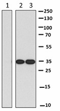
GFP-fused protein was immunoprecipitated from 100 µg transfected 293E cell extract using 10 µl anti-mCherry nanobody Affinity Gel (lane 1, negative control), 10 µl anti-GFP nanobody Affinity Gel (lane 2). Immunoprecipitates and 10% of total input (10 µg, Lane 3) were resolved by electrophoresis, transferred to nitrocellulose, and probed with purified anti-GFP (clone 1GFP63) antibody. Proteins were visualized using a goat anti-mouse-IgG secondary antibody conjugated to HRP and chemiluminescence detection.
Green fluorescent protein (GFP) was originally identified as a protein involved in bioluminescence, which is from the jellyfish Aequorea victoria. It is widely used as a fluorescent indicator for monitoring gene expression in a variety of cellular systems, including living organisms and fixed tissues. Unlike other bioluminescent reporters, GFP fluoresces without the need for exogenous substrates or cofactors, or other intrinsic or extrinsic proteins. This makes GFP a useful tool for monitoring gene expression and protein localization in vivo. Purified GFP is a 27 kD monomer consisting of 238 amino acids and emits green light (emission maximum at 509 nm) when excited with blue or UV light.
Nanobodies are heavy chain only, single-domain antibodies, that lack the presence of light chains. These are typically derived from members of the Camelidae family that include llamas and camels. Their variable region (VHH) is the smallest antigen-binding fragment found in a natural antibody, and nanobodies are the smallest (≈15 kD) naturally occuring immunoglobins. Further, nanobodies are stable, and can bind antigens with high affinity. Experimentally, nanobodies have been applied in WB, IF, and IP. In IP/WB applications, nanobodies can avoid IgG heavy/light chain issues that occur when using regular antibodies.
Product Details
- Verified Reactivity
- Aequorea victoria, Epitope tag
- Reported Reactivity
- Species independent
- Antibody Type
- Recombinant
- Immunogen
- Victoria GFP
- Formulation
- 50% anti-GFP nanobody (clone LaG-19) conjugated resin is supplied in 1X PBS and 0.09% NaN3. The volume specified for each catalog number indicates the volume of resin included.
- Preparation
- The antibody was purified by affinity chromatography.
- Storage & Handling
- Upon receipt, store between 2°C and 8°C. The unopened product is stable for one year upon arrival.
- Application
-
IP - Quality tested
- Recommended Usage
-
For immunoprecipitation, the suggested use of this reagent is 5 - 20 µl affinity gel for 100 µg lysate harvested from GFP overexpressed cells.
- RRID
-
AB_3105971 (BioLegend Cat. No. 689302)
AB_3105971 (BioLegend Cat. No. 689303)
AB_3105971 (BioLegend Cat. No. 689304)
Antigen Details
- Structure
- GFP is a 27 kD monomer consisting of 238 amino acids.
- Function
- Fluorescent protein.
- Biology Area
- Cell Biology
- Antigen References
-
1. Rizzuto R, et al. 1996. Curr. Biol. 6:183.
2. Chalfie M, et al. 1994. Science 263:802.
3. Cortez-Retamozo V, et al. 2004. Cancer Res. 64:2853.
4. Muyldermans S. 2013. Annu. Rev. Biochem. 82:775.
5. Fridy PC, et al. 2014. Nat. Methods 11:1253. - Gene ID
- NA
- UniProt
- View information about GFP on UniProt.org
Related Pages & Pathways
Pages
Related FAQs
Other Formats
View All GFP Reagents Request Custom Conjugation| Description | Clone | Applications |
|---|---|---|
| Anti-GFP Nanobody Affinity Gel | LaG-19 | IP |
Customers Also Purchased
Compare Data Across All Formats
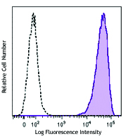
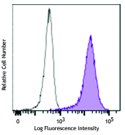
This data display is provided for general comparisons between formats.
Your actual data may vary due to variations in samples, target cells, instruments and their settings, staining conditions, and other factors.
If you need assistance with selecting the best format contact our expert technical support team.
-
Anti-GFP Nanobody Affinity Gel
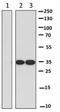
GFP-fused protein was immunoprecipitated from 100 µg transfe...




Follow Us