- Clone
- Poly18406 (See other available formats)
- Regulatory Status
- RUO
- Other Names
- Microtubule-Associated Protein 2, Map-2
- Previously
-
Covance Catalog# PRB-547C
- Isotype
- Rabbit Polyclonal IgG
- Ave. Rating
- Submit a Review
- Product Citations
- publications
-

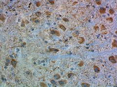
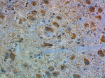
Figure Legend: IHC staining of purified anti-MAP2 antibody (Poly18406) on formalin-fixed, paraffin-embedded mouse brain. The tissue was incubated with primary antibody (1:2000 dilution) for 60 minutes at room temperature. BioLegend’s Ultra Streptavidin (USA) HRP Detection Kit (Multi-Species, DAB, Cat. No. 929501) was used for detection followed by hemotoxylin counterstaining, according to the protocol provided.
| Cat # | Size | Price | Save |
|---|---|---|---|
| 840601 | 100 µL |
Item is currently unavailable |
Microtubules are required for many well characterized functions in eukaryotic cells, including the movement of chromosomes in mitosis and meiosis, intracellular transport, establishment and maintenance of cellular morphology, cell growth, cell migration and morphogenesis in multicellular organisms. Microtubules are associated with a family of proteins called microtubule associated proteins (MAPs), which include the protein t (tau) and a group of proteins referred to as MAP1, MAP2, MAP3, MAP4 and MAP5. MAP2 is made up of two 280 kD apparent molecular weight bands referred to as MAP2a and MAP2b. A third lower molecular weight form, usually called MAP2c, corresponds to a pair of protein bands running at 70 kD on SDS-PAGE gels. All these MAP2 forms are derived from a single gene by alternate transcription.
Product DetailsProduct Details
- Verified Reactivity
- Mouse
- Reported Reactivity
- Other species
- Antibody Type
- Polyclonal
- Host Species
- Rabbit
- Immunogen
- The MAP2 antibody was raised using bovine brain MAP2, electrophoretically purified from heat stable MAP fraction and recognizes mammalian MAP2.
- Preparation
- Serum
- Storage & Handling
- Store at -20°C. Upon initial thawing, apportion into working aliquots and store at -20°C. Avoid repeated freeze-thaw cycles to prevent denaturing the antibody.
- Application
-
IHC-P - Quality tested
ICC - Reported in the literature, not verified in house - Recommended Usage
-
Each lot of this antibody is quality control tested by formalin-fixed paraffin-embedded immunohistochemical staining. For immunohistochemistry, a dilution range of 1:1000 - 1:2000 is suggested. It is recommended that the reagent be titrated for optimal performance for each application.
- Application Notes
-
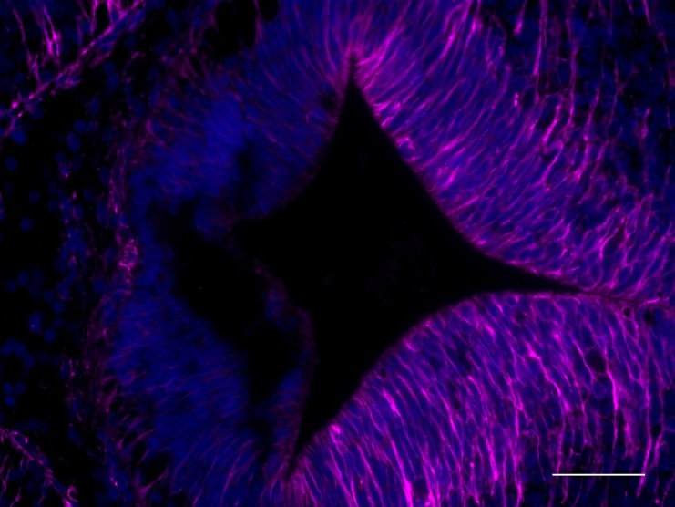
This antibody is effective in immunoblotting (WB), immunofluorescence (IF) and immunohistochemistry (IHC).
Positive control: Mouse brain
The antibody will stain neuronal cell bodies and dendritic processes, and will not stain glial cells or axons. It is the best marker to distinguish dendrites from axons in the brain.
This product may contain other non-IgG subtypes. -
Application References
(PubMed link indicates BioLegend citation) -
- De Camilli P, et al. 1984. Neuroscience. 11:817-46. (IHC)
- Bloom GS, et al. 1984. J Cell Biol. 98:320-30. (IHC)
- Maschietto M, et al. 2013. Front Cell Neurosci. 25:7. (IF) PubMed
- La Via L,et al. 2013. Nucelic Acids Res. 41:617. (IF) PubMed
- Ji B, et al. 2014. Hum Mol Genet. 23:5683-705. (ICC) PubMed
- Fu X, et al. 2013. J Neurosci. 33:709.
- Product Citations
-
- RRID
-
AB_2565455 (BioLegend Cat. No. 840601)
Antigen Details
- Biology Area
- Cell Biology, Neuroscience, Neuroscience Cell Markers
- Molecular Family
- Microtubules
- Gene ID
- 4133 View all products for this Gene ID
- UniProt
- View information about MAP2 on UniProt.org
Related FAQs
Other Formats
View All MAP2 Reagents Request Custom Conjugation| Description | Clone | Applications |
|---|---|---|
| Anti-MAP2 | Poly18406 | IHC-P,ICC |
Customers Also Purchased
Compare Data Across All Formats
This data display is provided for general comparisons between formats.
Your actual data may vary due to variations in samples, target cells, instruments and their settings, staining conditions, and other factors.
If you need assistance with selecting the best format contact our expert technical support team.




Follow Us