- Regulatory Status
- RUO
- Ave. Rating
- Submit a Review
- Product Citations
- publications
-

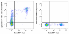
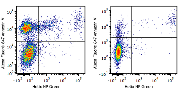
Heat induced 40% dead C57BL/6 splenocytes stained with Helix NP™ Blue and Apotracker™ Tetra Alexa Fluor® 647 (left) or Alexa Fluor® 647 Streptavidin (right). -

Extracellular vesicles (50-300 nm) were isolated from human plasma and stained with anti-human CD81 (clone 5A6) PE/Cyanine7 and Apotracker™ Tetra Alexa Fluor® 647 (left) or stained with anti-human CD81 (clone 5A6) PE/Cyanine7 only (right). Data shown was gated on events within the 100-250 nm size range. -

Extracellular vesicles (50-300 nm) were isolated from human plasma and stained with anti-human CD9 (clone HI9a) APC/Fire 750™ and Apotracker™ Tetra Alexa Fluor® 647 (left) or stained with anti-human CD9 (clone HI9a) APC/Fire 750™ only (right). Data shown was gated on events within the 100-250 nm size range.
| Cat # | Size | Price | Save |
|---|---|---|---|
| 427405 | 20 tests | ¥20,900 | |
| 427406 | 100 tests | ¥64,900 |
Apoptotic and/or necrotic cells display phosphatidylserine (PS) on their outer cell surface, where it acts as an ‘eat-me’ signal and contributes to efficient removal of dead cells by macrophages and other phagocytes. PS is normally only found on the intracellular leaflet of the plasma membrane in healthy cells, but during early apoptosis, membrane asymmetry is lost and PS translocates to the external leaflet. Apotracker™ monomer binds to PS for the detection and removal of dead or dying cells which are effective and preferably at the same time buffer-independent (Calcium free buffer) in comparison to the agents used in the market thus far.
Product DetailsKit Contents
- Kit Contents
-
For Cat# 427405
- 20 µL Apo-Monomer solution
- 24 µL Alexa Fluor® 647 Streptavidin solution
For Cat# 427406
- 100 µL Apo-Monomer solution
- 120 µL Alexa Fluor® 647 Streptavidin solution
Product Details
- Verified Reactivity
- Human, Mouse
- Formulation
-
Apo-Monomer: HEPES buffered saline w/ 8% Glycerol, pH 7.4
Streptavidin: Phosphate-buffered solution, pH 7.2, containing 0.09% sodium azide - Preparation
- The Apo monomer was purified by affinity chromatography and conjugated with biotin under optimal conditions. Streptavidin was conjugated with Alexa Fluor® 647 under optimal conditions.
- Storage & Handling
- Kit components should be stored undiluted between 2°C and 8°C, and protected from prolonged exposure to light. Do not freeze.
- Application
-
FC - Quality tested
- Recommended Usage
-
Each lot of this product is quality control tested by immunofluorescent staining with flow cytometric analysis. The suggested use of this reagent is 5 µL of the Apo-Monomer + Streptavidin-Fluorophore mixture per million cells in 100 µL volume.
Please see the detailed protocol in the application notes section below. - Excitation Laser
-
Red Laser (633 nm)
- Application Notes
-
Apotracker™ Tetra staining procedure for flow cytometry analysis:
-
To prepare 25 µL (5 tests) of the tetramer solution, mix 5 µL of Apo-monomer, 6 µL of Fluor-Streptavidin and 14 µL of Cell Staining Buffer (BioLegend Cat. No. 420201), or equivalent.
Note: We do not recommend preparing less than 25 µL (5 tests) of the Apo-monomer-Streptavidin complex at a time to avoid pipetting errors. Scale up as necessary
-
For each sample containing 1x10⁶ cells resuspended in 100 µL of Cell Staining Buffer, add 5 µL of tetramer solution prepared in the above step.
-
Incubate in the dark at room temperature for 15 minutes.
-
Wash each tube by adding 2 mL of Cell Staining Buffer, or equivalent, and centrifuging at 1200 rpm for five minutes. Remove the supernatant and vortex tubes to loosen pellet.
-
Repeat wash step above.
-
Resuspend the pellet by adding 300 - 500 μL Cell Staining Buffer to each tube.
-
(Optional) Ten minutes prior to flow cytometric analysis, add 10 μL of DAPI solution (or equivalent dye) to each tube for viability staining.
-
Cells are ready for flow cytometric analysis.
-
Antigen Details
- Gene ID
- NA




Follow Us