- Clone
- PY20 (See other available formats)
- Regulatory Status
- RUO
- Other Names
- p-Tyr
- Isotype
- Mouse IgG2b, κ
- Ave. Rating
- Submit a Review
- Product Citations
- publications
-

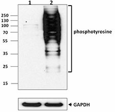
Western blot analysis of Hela untreated (lane 1) cell and Hela treated with Pervanadate for 15min (lane 2) using Biotin-anti-phosphotyrosine antibody (PY20). GAPDH antibody (poly6314) was used as loading control.
Phosphorylation is a common modification of proteins that can result in alterations in protein function, protein-protein association, cellular localization, and protein-half life. Phosphorylation can occur on threonine, serine, and tyrosine residues. The PY20 monoclonal antibody recognizes phosphorylated tyrosine residues in all species tested (human, mouse, rat, dog, chicken, and frog). The PY20 antibody has been shown to be useful for flow cytometry, immunoprecipitation, Western blotting, and immunofluorescence staining.
Product DetailsProduct Details
- Verified Reactivity
- Human, Mouse, Rat, All Species
- Antibody Type
- Monoclonal
- Host Species
- Mouse
- Immunogen
- KLH-conjugated phosphotyrosine
- Formulation
- Phosphate-buffered solution, pH 7.2, containing 0.09% sodium azide.
- Preparation
- The antibody was purified by affinity chromatography, and conjugated with biotin under optimal conditions.
- Concentration
- 0.5 mg/mL
- Storage & Handling
- The antibody solution should be stored undiluted between 2°C and 8°C. Do not freeze.
- Application
-
WB - Quality tested
- Recommended Usage
-
Each lot of this antibody is quality control tested by Western blotting. Suggested working dilution(s): Use 5 µg/5ml antibody dilution buffer per mini-gel. Do not use dilution or blocking buffers containing milk as they may interfere with antibody binding to proteins of interest. Dilution and blocking buffers containing 4% bovine serum albumin are recommended for use with this antibody. It is recommended that the reagent be titrated for optimal performance for each application.
- Application Notes
-
Additional reported applications (for the relevant formats) include: immunoprecipitation1,2, Western blotting1,2, immunocytochemistry3.
-
Application References
(PubMed link indicates BioLegend citation) -
- Vuori K, et al. 1995. J. Biol. Chem. 270:22259. (IP, WB)
- Glenney J, et al. 1988. J. Immunol. Meth. 109:277. (IP, WB)
- Prahalad P, et al. 2004. Am J Physiol Cell Physiol 286:C693. (IF)
- Zentillin L, et al. 2009. FASEB J. 24:1467. PubMed
- Philipsen L, et al. 2013. Mol Cell Proteomics. 12:2551. PubMed
- Cespedes PF, et al. 2014. PNAS. 111:3214. PubMed
- Product Citations
-
- RRID
-
AB_314766 (BioLegend Cat. No. 309304)
Antigen Details
- Distribution
-
Phospho-Specific
- Function
- Phosphorylation of specific tyrosine residues, signal transduction, cell cycle progression, oncogenic transformation
- Biology Area
- Cell Biology, Immunology, Signal Transduction
- Molecular Family
- Phospho-Proteins, Protein Kinases/Phosphatase
- Gene ID
- 5800 View all products for this Gene ID
- UniProt
- View information about Phosphotyrosine on UniProt.org
Related FAQs
- How many biotin molecules are per antibody structure?
- We don't routinely measure the number of biotins with our antibody products but the number of biotin molecules range from 3-6 molecules per antibody.
Other Formats
View All Phosphotyrosine Reagents Request Custom Conjugation| Description | Clone | Applications |
|---|---|---|
| Biotin anti-Phosphotyrosine | PY20 | WB |
| Purified anti-Phosphotyrosine | PY20 | WB,ICC,ICFC,IP |
| Alexa Fluor® 488 anti-Phosphotyrosine | PY20 | ICFC |
| Alexa Fluor® 647 anti-Phosphotyrosine | PY20 | ICFC |
| APC anti-Phosphotyrosine | PY20 | ICFC |
| PE anti-Phosphotyrosine | PY20 | ICFC |
| Alexa Fluor® 594 anti-Phosphotyrosine | PY20 | ICC |
Customers Also Purchased
Compare Data Across All Formats
This data display is provided for general comparisons between formats.
Your actual data may vary due to variations in samples, target cells, instruments and their settings, staining conditions, and other factors.
If you need assistance with selecting the best format contact our expert technical support team.
-
Biotin anti-Phosphotyrosine
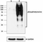
Western blot analysis of Hela untreated (lane 1) cell and He... -
Purified anti-Phosphotyrosine
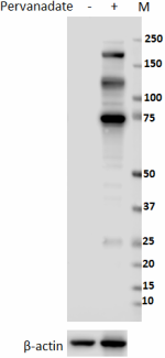
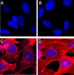
Total cell lysate (15 µg protein) from non-treated and 1mM P... NIH3T3 cells were non-treated (A and B) or treated with 1mM ... -
Alexa Fluor® 488 anti-Phosphotyrosine
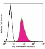
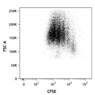
Hydrogen peroxide stimulated EL4 cells intracellularly stain... -
Alexa Fluor® 647 anti-Phosphotyrosine
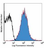
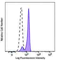
Hydrogen peroxide stimulated EL4 cells intracellularly stain... -
APC anti-Phosphotyrosine
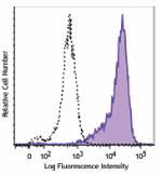
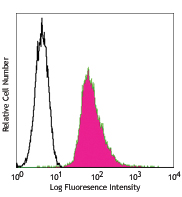
Human T leukemia cell line Jurkat was cultured in serum-free... -
PE anti-Phosphotyrosine
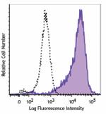
Human T leukemia cell line Jurkat was cultured in serum-free... -
Alexa Fluor® 594 anti-Phosphotyrosine
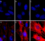
NIH3T3 cells were non-treated (A-C) or treated with 1mM perv...





Follow Us