- Regulatory Status
- RUO
- Other Names
- Live/Dead Staining, Dead cell exclusion dye, Cell viability dye
- Ave. Rating
- Submit a Review
- Product Citations
- publications

-

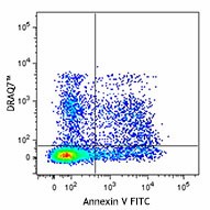
C57BL/6 mouse splenocytes were stored at 4°C for 1 day and then stained with Annexin V FITC, washed, and then stained with DRAQ7™ (1:1000). Cells were acquired on a 633 nm laser equipped-flow cytometer.
DRAQ7™ is a far-red emitting, anthraquinone compound that stains nuclei in dead and permeablized cells. It is non-permeant to live cells and thus can be used for differentiating live and dead cells. It is optimally excited at 633 nm, and can be detected using 695LP, 715LP, and 780LP filters. DRAQ7™ can also be used in cell imaging as it does not get excited by UV or Violet lasers and is sub-optimally excited by the 488 nm laser. It can be combined with FITC, PE, and other UV or violet excitable dyes for multi-color analysis. DRAQ7™ is non-toxic and thus can be used for siRNA studies and other dynamic viablity assays.
Product DetailsProduct Details
- Preparation
- DRAQ7™ is supplied at 1 ml per vial (300 µM).
- Storage & Handling
- DRAQ7™ should be stored between 2°C and 8°C upon receipt.
- Application
-
FC - Quality tested
- Recommended Usage
-
DRAQ7™ is a trademark of Biostatus Limited.
- Application Notes
-
DRAQ7™ is a membrane impermeable dye that only stains the nuclei of dead or permeabilized cells. It is a far-red emitting dye with optimal excitation/ emission peaks of 633nm/ 695nm, respectively. Suboptimally, DRAQ7™ can be excited at 561nm and 594nm as well, with the same emission peak occurring at 695nm. Ideally, this probe should be detected in the Alexa Fluor® 700 and APC/Cyanine7 emission filters off of the 633nm laser.
Protocol for Dead Cell/Apoptosis Analysis using DRAQ7™:
1. Perform surface staining or Annexin V staining following protocol of choice.
2. Wash cells twice with phosphate buffered saline (PBS). Sodium azide interferes with DRAQ7™ staining; therefore it is recommended to stain in PBS (without calcium or magnesium) or in culture medium without sodium azide.
3. Dilute DRAQ7™ to your required concentration. We recommend titrating the reagent to determine optimal concentration for cells of interest. Good results have been observed using a 1:200 to 1:1000 dilution.
4. Incubate at room temperature, preventing exposure to light, for 10-15 minutes. No washing is necessary.
5. Analyze cells on a cytometer equipped with the appropriate laser. - Product Citations
-
Antigen Details
- Biology Area
- Cell Biology, Cell Cycle/DNA Replication, Cell Motility/Cytoskeleton/Structure, Cell Proliferation and Viability, Neuroscience
- Antigen References
-
1. Akagi J, et al. 2013. Cytometry A. 83: 227.
2. Smith PJ, et al. 2013. Cytometry A. 659-71.
3. Wlodokowic D, et al. 2013. Curr Protoc Cytom. Chapter 9. Unit 9.4.1 - Gene ID
- NA
Related Pages & Pathways
Pages
Related FAQs
Customers Also Purchased


Follow Us