


- Regulatory Status
- RUO
- Other Names
- FOXP3 Fixation/Permeabilization Buffer
- Ave. Rating
- Submit a Review
- Product Citations
- publications

-

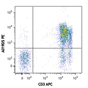
Whole blood was stained with human CD3 APC/Cyanine7 (blue and red) or APC/Fire™ 750 (green and purple), then lysed, washed and fixed under the following conditions: A) Fixed with FOXP3 fix buffer followed by permeabilization with FOXP3 perm buffer (purple and red). B) Fixed with True-Nuclear™ fix buffer, then permeabilized with True-Nuclear™ perm buffer (green and blue). Compensation requirements between the APC and APC/Cyanine7 channels increased significantly for the FoxP3 Fix/Perm buffer set versus the True-Nuclear™ transcription factor buffer set for both APC/Cyanine7 and APC/Fire™ 750. -

BALB/c spleen cells were cultured for four days in presence of LPS. Then the cells were stained with CD45R/B220 PE, followed by fixation and permeabilization with either FoxP3 Fix/Perm Buffer (top) or True-Nuclear™ Transcription Factor Buffer Set (bottom), and staining with Blimp-1 (clone 5E7) Alexa Fluor® 647. Data shown was gated on live cells using Zombie Aqua™ fixable viability dye. -

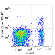
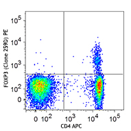
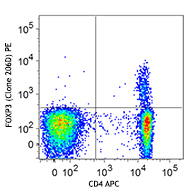
Human peripheral blood lymphocytes were surface stained with CD4 APC and then treated with True-Nuclear™ Transcription Factor Buffer Set (Cat. No. 424401). Cells were then stained with FOXP3 (clone 206D) Brilliant Violet 421™ (top) or mouse IgG1, κ Brilliant Violet 421™ isotype control (bottom).
BioLegend's FOXP3 Fix/Perm Buffer Set has been specially formulated for intracellular staining FOXP3 with minimum effect on the surface fluorochrome staining. It contains one 30 ml 4X concentrated FOXP3 Fix/Perm buffer (Cat. No. 421401) and one 25 mL 10X concentrated FOXP3 Perm buffer (Cat. No. 421402). The FOXP3 Fix/Perm buffer (4X) must be freshly diluted to 1X working solution with PBS (10 mM sodium phosphate, 150 mM sodium chloride, pH 7.2) prior to staining procedures. e.g. Dilute one (1) part FOXP3 Fix/Perm buffer (4X) to three (3) parts PBS. The FOXP3 Perm buffer (10X) should be diluted to 1X working solution with PBS (10 mM sodium phosphate, 150 mM sodium chloride, pH 7.2) prior to staining procedures. e.g. Dilute one (1) part FOXP3 Perm buffer(10X) to nine (9) parts PBS.
Warning: The FOXP3 Buffer Set can have a deleterious effect on tandem fluorophores (particularly APC/Cyanine7) in a multicolor assay, causing an increase in compensation into the APC channel. If your panel contains a surface stain with an antibody conjugated to a tandem fluorophore, consider using the True-Nuclear™ Transcription Factor Buffer Set (Cat. No. 424401) instead, which provides a clearer resolution of FoxP3 as well as other nuclear transcription factors. Please refer to the data provided here.
Caution: The FOXP3 Fix/Perm buffer contains paraformaldehyde, which is toxigenic and mutagenic. Please handle with caution and wear gloves, lab coat and necessary protection to avoid direct body contact.
Product Details
- Storage & Handling
-
Store between 2°C and 8°C. Do not freeze.To obtain lot-specific expiration date, please enter the lot number in our Concentration and Expiration Lookup or Certificate of Analysis online tools.)
This product has a shelf-life of 12 months or less. Please contact our technical support team for lot specific CoA and expiration date inquiries of this product. - Application
-
ICFC - Quality tested
- Recommended Usage
-
The FOXP3 Fix/Perm buffer(4X) must be diluted to 1X working solution with PBS (10 mM sodium phosphate, 150 mM sodium chloride, pH 7.2) prior to staining procedures. e.g. Dilute one (1) part FOXP3 Fix/Perm buffer(4X) to three (3) parts PBS. The FOXP3 Perm buffer (10X) should be diluted to 1X working solution with PBS (10 mM sodium phosphate, 150 mM sodium chloride, pH 7.2) prior to staining procedures. e.g. Dilute one (1) part FOXP3 Perm buffer(10X) to nine (9) parts PBS.
NOTE: The FOXP3 Perm buffer (10X) may have crystalization or precipitation oberserved when it is stored at 2-8°C, however, it's normal and does not affect the buffer performance. If there is a heavy precipitation observed after diluted to 1X working solution, it can be filtered to clarify the solution. - Application Notes
-
NOTE: For flow cytometric staining, True-Nuclear™ Transcription Factor Buffer Set (Cat. No. 424401) offers improved staining and is highly recommended over the Foxp3 Fix/Perm Buffer Set (Cat. No. 421403).
FOXP3 Intracellular Staining Procedures:
1. Perform cell surface staining as described in BioLegend's Cell Surface Immunofluorescence Staining Protocol.
2. Add 1 ml of 1X BioLegend's FOXP3 Fix/Perm solution to each tube, vortex and incubate at room temperature in the dark for 20 minutes, then spin down the cells and remove the supernatant.
3. Wash once with cell staining buffer (Cat. No. 420201) by spin at 250Xg for 5 minutes and remove the supernatant.
4. Wash once with 1ml 1X BioLegend's FOXP3 Perm buffer.
5. Re-suspend cells in 1ml 1X BioLegend's FOXP3 Perm buffer, incubate at room temperature in the dark for 15 minutes, spin down cells and discard the supernatant, then resuspend the pellet in 100 ul of 1X BioLegend's FOXP3 Perm buffer.
6. Add appropriate amount of flurochrome conjugated anti-FOXP3 antibody and incubate at room temperature in the dark for 30 minutes.
7. Wash twice with cell staining buffer, and resuspend in 0.5ml cell staining buffer then analyze with flow cytometer with appropriate instrument setting. -
Application References
(PubMed link indicates BioLegend citation) -
- Yang ZZ, et al. 2006. Blood 107:3639. PubMed
- Groh V, et al. 2006. Nat. Immunol. 7:755. PubMed
- Bamias G, et al. 2007. J. Immunol. 178:1809. PubMed
- MacDonald K PA, et al. 2007. Blood doi:10.1182/blood-2007-01-067249. PubMed
- Müller M, et al. 2007. J. Immunol. 179:2774.
- Dang Y,et al.2007.Clin Cancer Res.13:1883. PubMed
- Basu S, et al. 2008. J. Immunol. 180:5974. PubMed
- Takamura S, et al. 2010. J. Immunol. 184:4696. PubMed
- Beavis, PA., et al. 2011. PNAS. 108:16717. PubMed
- Kling J, et al. 2013. Exp Parasitol. 129:270. PubMed
- Troutman TD, et al. 2012. PNAS. 109:273. PubMed
- Faustman DL, et al. 2012. PLoS One. 7:e41756. PubMed
- Liu Y, et al. 2012. Food Chem Toxicol. 50:1920. PubMed
- Kawamoto N, et al. 2013. Int Immunol. 25:53. PubMed
- Morandi B, et al. 2013. J. Immunol. 191:4858. PubMed
- Shiba T, et al. 2014. Toxicol Appl Pharmacol. 274:191. PubMed
- Cook KW, et al. 2014. Gut. PubMed
- Shibuya KC, et al. 2014. PLoS One. 9:96565. PubMed
- Cecil DL, et al. 2014. Cancer Res. 74:2710. PubMed
- Linnerbauer S, et al. 2014. PLoS Pathog. 10:1004068. PubMed
- Basu S, et al. 2015. J Leukoc Biol. 97:279. PubMed
- Product Citations
-
Antigen Details
- Cell Type
- Tregs
- Biology Area
- Immunology, Transcription Factors
- Molecular Family
- Nuclear Markers
- Gene ID
- NA


Related FAQs
- Can I stain whole blood with anti-FOXP3 using your Foxp3 staining kit?
-
It is not recommended. It is best to use PBMCs for this testing.
- Can FOXP3 be costained with cytokines?
-
The larger holes created by the nuclear permeabilization required for FOXP3 may allow cytokines to leak out of the cell, making it harder to detect lowly-expressed cytokines. You may have to use a control where the cells are only permeabilized through the cell membrane.
Customers Also Purchased


Follow Us