- Clone
- W7C6 (See other available formats)
- Regulatory Status
- RUO
- Other Names
- Mesenchymal Stem Cells (MSC), Leukocyte Associated Antigen (LAR)
- Isotype
- Mouse IgG1, κ
- Ave. Rating
- Submit a Review
- Product Citations
- publications

-

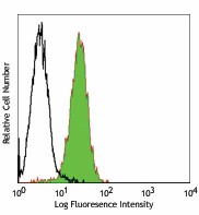
Human cervical cancer cell line, HeLa, stained with W7C6 PE -

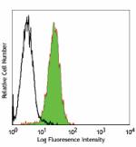
Human bone marrow-derived mesenchymal stem cells stained with W7C6 PE
The W7C6 antibody was generated by immunization with Weri-RB-1 cells. It reacts with mesenchymal stem cells and some tested neoplastic cell lines (HT29, Hela, MCF-7, REH). Further studies have shown that W7C6 antigen is identical to protein tyrosine phosphatase LAR. Mesenchymal stem cells or MSCs are originally name as bone marrow stromal cells. They are a population of adult stem cells with a large capacity for self-renewal and multipotency for differentiation into a variety of cell types including osteoblasts, chondrocytes, myocytes, adipocytes, β-pancreatic islets cells and certain neuronal cells. MSCs reside in many tissues, such as bone marrow, placenta, adipose tissue, adult peripheral blood, fetal blood, skin, as well as liver and lung. Besides their plasticity for tissue repair, MSCs also exhibit a powerful immunosuppressive activity and play important role in supporting hematopoiesis.
Product DetailsProduct Details
- Verified Reactivity
- Human
- Reported Reactivity
- African Green, Baboon, Cynomolgus, Rhesus
- Antibody Type
- Monoclonal
- Host Species
- Mouse
- Immunogen
- Retinoblastoma cell line (Weri-RB-1 cells)
- Formulation
- Phosphate-buffered solution, pH 7.2, containing 0.09% sodium azide and BSA (origin USA)
- Preparation
- The antibody was purified by affinity chromatography, and conjugated with PE under optimal conditions.
- Concentration
- Lot-specific (to obtain lot-specific concentration and expiration, please enter the lot number in our Certificate of Analysis online tool.)
- Storage & Handling
- The antibody solution should be stored undiluted between 2°C and 8°C, and protected from prolonged exposure to light. Do not freeze.
- Application
-
FC - Quality tested
- Recommended Usage
-
Each lot of this antibody is quality control tested by immunofluorescent staining with flow cytometric analysis. For flow cytometric staining, the suggested use of this reagent is 5 µl per million cells in 100 µl staining volume or 5 µl per 100 µl of whole blood.
- Excitation Laser
-
Blue Laser (488 nm)
Green Laser (532 nm)/Yellow-Green Laser (561 nm)
- RRID
-
AB_2266513 (BioLegend Cat. No. 327606)
Antigen Details
- Distribution
-
Mesenchymal stem cells, some neoplastic cell lines (HT29, Hela, MCF-7, REH)
- Cell Type
- Mesenchymal cells
- Biology Area
- Immunology
- Antigen References
-
1. Buhring HJ, et al. 2007. Ann. N. Y. Acad. Sci. 1106:262.
2. Jackson L, et al. 2007 J. Postgrad. Med. 53:121.
3. Krampera M, et al. 2003 Blood 101:3722.
4. Ball LM, et al. 2007 Blood 110:2764 - Gene ID
- NA
- UniProt
- View information about MSC on UniProt.org
Related Pages & Pathways
Pages
Related FAQs
- What type of PE do you use in your conjugates?
- We use R-PE in our conjugates.
Other Formats
View All MSC Reagents Request Custom Conjugation| Description | Clone | Applications |
|---|---|---|
| PE anti-human MSC (W7C6) | W7C6 | FC |
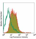
Compare Data Across All Formats
This data display is provided for general comparisons between formats.
Your actual data may vary due to variations in samples, target cells, instruments and their settings, staining conditions, and other factors.
If you need assistance with selecting the best format contact our expert technical support team.


Follow Us