- Clone
- 5E3/SEMA4A (See other available formats)
- Regulatory Status
- RUO
- Other Names
- Semaphorin 4A, SEMAB, Sema B, Semaphorin B, RP35, CORD10, SEMA 4A
- Isotype
- Mouse IgG1, λ
- Ave. Rating
- Submit a Review
- Product Citations
- publications
-

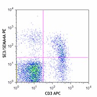
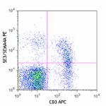
Mouse splenocytes were stained with CD3 APC and Semaphorin 4A (clone 5E3/SEMA4A) PE (top) or mouse IgG1 PE isotype control (bottom). -

| Cat # | Size | Price | Save |
|---|---|---|---|
| 148403 | 25 µg | ¥28,380 | |
| 148404 | 100 µg | ¥64,240 |
Semaphorin 4A (SEMA4A) is a class IV semaphorin of soluble and transmembrane proteins originally identified as SemB. It is a single-pass type I membrane protein containing an immunoglobulin-like C2-type domain, a PSI domain, and a Sema domain. SEMA4A is expressed on dendritic cells, activated B cells, and T cells. It is also found on different tissues, such as those in brain, lung, kindney, spleen, and testis. In the immune system, SEMA4A functions as an activator in the T-cell-mediated immune response and suppresses vascular endothelial growth factor (VEGF)-mediated endothelial cell migration and proliferation in vitro and angiogenesis in vivo. Like other semaphorin family members it is also involved in guidance of axonal migration during neuronal development and in immune responses.
Product DetailsProduct Details
- Verified Reactivity
- Mouse, Human
- Antibody Type
- Monoclonal
- Host Species
- Mouse
- Immunogen
- Mouse SEMA4A-Fc proteins
- Formulation
- Phosphate-buffered solution, pH 7.2, containing 0.09% sodium azide.
- Preparation
- The antibody was purified by affinity chromatography and conjugated with PE under optimal conditions.
- Concentration
- 0.2 mg/ml
- Storage & Handling
- The antibody solution should be stored undiluted between 2°C and 8°C, and protected from prolonged exposure to light. Do not freeze.
- Application
-
FC - Quality tested
- Recommended Usage
-
Each lot of this antibody is quality control tested by immunofluorescent staining with flow cytometric analysis. For flow cytometric staining, the suggested use of this reagent is ≤0.25 µg per million cells in 100 µl volume. It is recommended that the reagent be titrated for optimal performance for each application.
- Excitation Laser
-
Blue Laser (488 nm)
Green Laser (532 nm)/Yellow-Green Laser (561 nm)
- Application Notes
-
Additional reported applications (for relevant formats) include: ELISA1.
-
Application References
(PubMed link indicates BioLegend citation) -
- Kumanogoh A. et al. 2005. Immunity. 22:305. (ELISA)
- Product Citations
-
- RRID
-
AB_2565286 (BioLegend Cat. No. 148403)
AB_2565286 (BioLegend Cat. No. 148404)
Antigen Details
- Structure
- Single-pass type I membrane protein containing an immunoglobulin-like C2-type domain, a PSI domain and a SEMA domain.
- Distribution
-
Dendritic cells, T cells, B cells, brain, lung, kindney, spleen and testis
- Function
- An activator in T-cell-mediated immune response. Suppresses vascular endothelial growth factor (VEGF)-mediated endothelial cell migration and proliferation in vitro and angiogenesis in vivo. It is also involved in guidance of axonal migration during neuronal development.
- Cell Type
- Dendritic cells, T cells, B cells, Tregs
- Biology Area
- Cell Biology, Immunology, Innate Immunity, Neuroscience, Synaptic Biology
- Molecular Family
- Adhesion Molecules
- Antigen References
-
1. Kumanogoh A, et al. 2002. Nature. 419:629.
2. Kumanogoh A, et al. 2005. Immunity. 22:305.
3. Toyofuku T, et al. 2007. EMBO J. 26:1373.
4. Delgoffe GM, et al. 2013. Nature. 501:252. - Gene ID
- 20351 View all products for this Gene ID 64218 View all products for this Gene ID
- UniProt
- View information about SEMA4A on UniProt.org
Related Pages & Pathways
Pages
Related FAQs
- What type of PE do you use in your conjugates?
- We use R-PE in our conjugates.
Other Formats
View All SEMA4A Reagents Request Custom Conjugation| Description | Clone | Applications |
|---|---|---|
| Purified anti-mouse/human SEMA4A | 5E3/SEMA4A | FC,ELISA |
| PE anti-mouse/human SEMA4A | 5E3/SEMA4A | FC |
| APC anti-mouse/human SEMA4A | 5E3/SEMA4A | FC |
Customers Also Purchased
Compare Data Across All Formats
This data display is provided for general comparisons between formats.
Your actual data may vary due to variations in samples, target cells, instruments and their settings, staining conditions, and other factors.
If you need assistance with selecting the best format contact our expert technical support team.
-
Purified anti-mouse/human SEMA4A
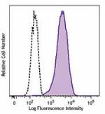
Human peripheral blood monocyte derived dendritic cells were... 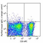
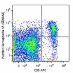
C57BL/6 mouse splenocytes were stained with CD3 APC and puri... -
PE anti-mouse/human SEMA4A
Mouse splenocytes were stained with CD3 APC and Semaphorin 4... 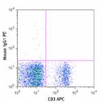
-
APC anti-mouse/human SEMA4A

Mouse splenocytes were stained with CD3 Pacific Blue™ and Se...






Follow Us