- Clone
- M1401E03 (See other available formats)
- Regulatory Status
- RUO
- Other Names
- Epidermal Growth Factor, Urogastrone (URG), HOMG4
- Isotype
- Mouse IgG1, κ
- Ave. Rating
- Submit a Review
- Product Citations
- publications
-
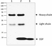
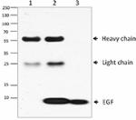
Immunoprecipitation/Western blot analysis of 200ng recombinant EGF. Lane 1 was immunoprecipitated with mouse IgG control antibody, lane 2 was immunoprecipitated with EGF antibody (clone M1401E03), and lane 3 was loaded with 25ng recombinant human EGF. Western blot analysis was performed using goat anti-human EGF antibody followed by HRP-anti-goat IgG.
Epidermal growth factor (EGF) is a small 6 kD polypeptide and has six conserved cysteine residues that form three intramolecular disulfide bonds. Human EGF is synthesized as transmembrane precursor proteins (1207 amino acids), which are proteolytically cleaved to generate the 54 amino acid mature EGF. EGF/EGF receptor signaling pathway plays an important role in proliferation, differentiation, and migration of a variety of cell types, especially in epithelial cells. The binding of EGF to EGFR induces receptor dimerization, which is required for activating the tyrosine kinase dimerization in the receptor cytoplasmic domain. In addition, the binding of EGF to its receptor triggers several signal transduction pathways including JAK/STAT, Ras/ERK/, and PI3K/AKT pathways. The blocking of the EGF/EGFR pathway can suppress some tumor cell proliferation.
Product DetailsProduct Details
- Verified Reactivity
- Human
- Antibody Type
- Monoclonal
- Host Species
- Mouse
- Immunogen
- Human EGF, amino acids Asn971 - Arg1023, with an N-terminal Met (Accession# P01133) was expressed in E. coli.
- Formulation
- Phosphate-buffered solution, pH 7.2, containing 0.09% sodium azide.
- Preparation
- The antibody was purified by affinity chromatography.
- Concentration
- 0.5 mg/ml
- Storage & Handling
- The antibody solution should be stored undiluted between 2°C and 8°C.
- Application
-
IP - Quality tested
- Recommended Usage
-
Each lot of this antibody is quality control tested by immunoprecipitation. For immunoprecipitation, the suggested use of this reagent is 2-10 µg per ml. It is recommended that the reagent be titrated for optimal performance for each application.
- Product Citations
-
- RRID
-
AB_2566190 (BioLegend Cat. No. 679502)
Antigen Details
- Structure
- 54 amino acids wtih a predicted molecular weight of approximately 6 kD.
- Distribution
-
Mammary gland cells, macrophages, gut epithelial cells, cells in the nervous system, and the kidney.
- Function
- EGF is a potent mitogen for many cells in culture, and in vivo, it induces the proliferation and the differentiation of the skin, cornea, lungs, trachea, and among other tissues. Processing of pro EGF in different tissues is not equally efficient. The precursor is processed to mature EGF in the submaxillary glands, pancreas, small intestine, and the mammary glands. EGF is fully processed, stored in secretory granules, and secreted in saliva. In the kidney, EGF is present in unprocessed or intermediated forms on the cell surface.
- Interaction
- EGFR, RHBDF2, RHBDF1, and ERAD.
- Ligand/Receptor
- EGFR.
- Biology Area
- Cell Biology, Cell Cycle/DNA Replication, Immunology, Signal Transduction
- Molecular Family
- Cytokines/Chemokines, Growth Factors
- Antigen References
-
1. Henson ES and Gibson SB. 2006. Cell Signal. 18:2089.
2. Burgess AW, et al. 2003. Mol. Cell. 12:541.
3. Imai Y, et al. 1982. Cancer Res. 42:4394.
4. Barnes DW. 1982. J. Cell. Biol. 93:1.
5. Heo JS, et al. 2006. Am. J. Physiol. Cell Physiol. 290:C123.
6. Bodnar RJ. 2013. Adv. Wound Care (New Rochelle). 2:24.
7. Zeng F, et al. 2014. Semin. Cell Dev. Biol. 28:2. - Gene ID
- 1950 View all products for this Gene ID
- UniProt
- View information about EGF on UniProt.org
Related FAQs
Other Formats
View All EGF Reagents Request Custom Conjugation| Description | Clone | Applications |
|---|---|---|
| Purified anti-EGF | M1401E03 | IP |
Customers Also Purchased
Compare Data Across All Formats
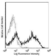
This data display is provided for general comparisons between formats.
Your actual data may vary due to variations in samples, target cells, instruments and their settings, staining conditions, and other factors.
If you need assistance with selecting the best format contact our expert technical support team.
-
Purified anti-EGF
Immunoprecipitation/Western blot analysis of 200ng recombina...




Follow Us