- Clone
- 5E10-D8 (See other available formats)
- Regulatory Status
- RUO
- Other Names
- histone H4, histone 1, H4a, H4 histone family member A
- Previously
-
Covance Catalog# MMS-5286
- Isotype
- Mouse IgG1, κ
- Ave. Rating
- Submit a Review
- Product Citations
- publications
-

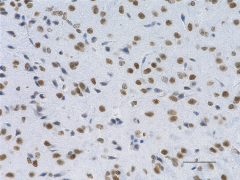
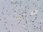
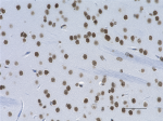
IHC staining of purified anti-Histone H4 Monomethyl (Lys20) antibody (clone 5E10-D8) on formalin-fixed paraffin-embedded mouse brain tissue. Following antigen retrieval using Sodium Citrate H.I.E.R., the tissue was incubated with 10 µg/ml of the primary antibody overnight at 4°C. BioLegend's Ultra-Streptavidin (USA) HRP kit (Multi-Species, DAB, Cat. No. 929901) was used for detection followed by hematoxylin counterstaining, according to the protocol provided. The image was captured with a 40X objective. Scale bar: 50 µm -

IHC staining of purified anti-Histone H4 Monomethyl (Lys20) antibody (clone 5E10-D8) on formalin-fixed paraffin-embedded human brain tissue. Following antigen retrieval using Sodium Citrate H.I.E.R., the tissue was incubated with 5 µg/ml of the primary antibody overnight at 4°C. BioLegend's Ultra-Streptavidin (USA) HRP kit (Multi-Species, DAB, Cat. No. 929901) was used for detection followed by hematoxylin counterstaining, according to the protocol provided. The image was captured with a 40X objective. Scale bar: 50 µm -

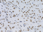
IHC staining of purified anti-Histone H4 Monomethyl (Lys20) antibody (clone 5E10-D8) on formalin-fixed paraffin-embedded rat brain tissue. Following antigen retrieval using Sodium Citrate H.I.E.R., the tissue was incubated with 5 µg/ml of the primary antibody overnight at 4°C. BioLegend's Ultra-Streptavidin (USA) HRP kit (Multi-Species, DAB, Cat. No. 929901) was used for detection followed by hematoxylin counterstaining, according to the protocol provided. The image was captured with a 40X objective. Scale bar: 50 µm -

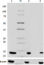
Total lysates (15µg protein) from Jurkat (lane1), Raw 264.7 (lane 2) and UMR106 (lane 3) cells were resolved by electrophoresis (4-20% Tris-Glycine gel), transferred to nitrocellulose, and probed with 1:5000 diluted (0.1 µg/mL) Purified anti-Histone H4 Monomethyl (Lys20) Antibody, clone 5E10-D8 (upper). Proteins were visualized by chemiluminescence detection using a 1:3000 diluted anti-Mouse-IgG secondary antibody conjugated to HRP for the anti-Histone H4 Monomethyl (Lys20) Antibody or 1:5000 diluted Direct-Blot™ HRP anti-β-Actin Antibody, clone 2F1-1(lower). Lane M: Molecular weight ladder.
Histone proteins are classified into core histones (H2A, H2B, H3, H4) and linker histones (H1, H5). Core histones form an octamer, which contains two H2A-H2B dimers and one H3-H4 tetramer. Core histones are predominantly globular except for the unstructured N-terminal tails. Posttranslational modifications, such as acetylation, methylation, phosphorylation, ubiquitination, SUMOylation and ADP-ribosylation occur in histone tails.
Histone modifications induce changes of chromatin structure and thereby affect the accessibility of transcription factors, nuclear proteins and enzymes to genomic DNA, resulting in gene activation or repression. It is known that histone modifications play critical roles in DNA repair, DNA replication, transcription regulation, alternative splicing and chromosome condensation and some diseases including autoimmune diseases and cancers.
Product Details
- Verified Reactivity
- Human, Rat, Mouse
- Antibody Type
- Monoclonal
- Host Species
- Mouse
- Immunogen
- This monoclonal antibody was raised against a synthetic peptide conjugated to KLH containing methylated lysine 20 of human Histone H4.
- Formulation
- Phosphate-buffered solution.
- Preparation
- The antibody was purified by affinity chromatography.
- Concentration
- 1.0 mg/ml
- Storage & Handling
- This antibody should be handled aseptically as it is free of preservatives such as Sodium Azide. Store this antibody undiluted between 2°C and 8°C. Please note the storage condition for this antibody has been changed from -20°C to between 2°C and 8°C. You can also check the vial label or CoA to find the proper storage conditions.
- Application
-
IHC-P - Quality tested
WB - Verified - Recommended Usage
-
Each lot of this antibody is quality control tested by formalin-fixed paraffin-embedded immunohistochemical staining. For immunohistochemistry, a concentration range of 5 - 10 µg/ml is suggested. For Western blotting, the suggested use of this reagent is 0.1 - 1 µg per ml. It is recommended that the reagent be titrated for optimal performance for each application.
-
Application References
(PubMed link indicates BioLegend citation) -
- Shirakata Y, et al. 2014. J. Reprod. Dev. 60:383. (IHC-P)
- RRID
-
AB_2564916 (BioLegend Cat. No. 828001)
Antigen Details
- Structure
- Histone H4 is a 103 amino acid protein with an apparent molecular mass of 12 kD
- Cell Type
- Mesenchymal Stem Cells
- Biology Area
- Cell Biology, Neuroscience, Stem Cells
- Gene ID
- 8359 View all products for this Gene ID
- UniProt
- View information about Histone on UniProt.org
Related Pages & Pathways
Pages
Other Formats
View All Histone Reagents Request Custom Conjugation| Description | Clone | Applications |
|---|---|---|
| Purified anti-Histone H4 Monomethyl (Lys20) | 5E10-D8 | IHC-P,WB |
Customers Also Purchased
Compare Data Across All Formats
This data display is provided for general comparisons between formats.
Your actual data may vary due to variations in samples, target cells, instruments and their settings, staining conditions, and other factors.
If you need assistance with selecting the best format contact our expert technical support team.




Follow Us