- Clone
- 3E8 (See other available formats)
- Regulatory Status
- RUO
- Other Names
- High mobility group protein 1, High mobility group protein B1, Sulfoglucuronyl carbohydrate binding protein
- Isotype
- Mouse IgG2b, κ
- Ave. Rating
- Submit a Review
- Product Citations
- publications
-

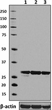
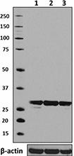
Total cell lysate from HeLa (lane 1, 15 µg), Raw264.7 (lane 2, 15 µg) and HepG2 (lane 3, 15 µg) were resolved by electrophoresis (4-12% Bis-Tris), transferred to nitrocellulose, and probed with 1µg/ml purified anti-HMGB1 antibody (clone 3E8). Proteins were visualized using an HRP Goat anti-mouse IgG Antibody and chemiluminescence detection. Direct-Blot™ HRP anti-β-actin antibody (clone 2F1-1) was used as a loading control. -

HeLa cells were fixed with 2% PFA for ten minutes, permeabilized with 0.5% Triton X-100 for five minutes, and blocked with 5% FBS for 30 minutes. Then the cells were intracellularly stained with 2 µg/mL anti-HMGB1 Antibody (clone 3E8) followed by Alexa Fluor® 594 (red) conjugated goat anti-mouse IgG in blocking buffer for two hours at room temperature. Actin filaments were labeled with Alexa Fluor® 488 Phalloidin (green). Nuclei were counterstained with DAPI (blue). The image was captured with a 60X objective. -

IHC staining using purified anti-HMGB1 (clone 3E8) on formalin-fixed paraffin-embedded human tonsil tissue. Following antigen retrieval using 1X Tris-Buffered Saline (final concentration 0.05 M) with Tween-20 (Cat. No. 925501), the tissue was incubated with 10 μg/mL of antibody overnight at 4°C, followed by incubation with 2.5 μg/mL of Alexa Fluor® 647 goat anti-mouse IgG antibody (Cat. No. 405322) for one hour at room temperature. Nuclei were counterstained with DAPI (blue) (Cat. No. 422801), and the slide was mounted with ProLong™ Gold Antifade Mountant. The image was captured with a 40X objective. Scalebar = 50 μM
| Cat # | Size | Price | Save |
|---|---|---|---|
| 651401 | 25 µg | ¥32,340 | |
| 651402 | 100 µg | ¥71,060 |
High mobility group protein B1 (HMGB1) belongs to a family of highly conserved proteins that contain HMG box domains. Human HMGB1 is expressed as a 215 amino acid (aa) single chain polypeptide containing three domains: two N-terminal globular, 70 aa positively charged DNA binding domains (HMG boxes A and B), and a negatively charged 30 aa C-terminal region that contains only Asp and Glu. Human HMGB1 is 100% aa identical to canine HMGB1 and 99% aa identical to mouse, rat, bovine and porcine HMGB1.
HMGB1 is a widely expressed and highly abundant protein. It was originally discovered as a nuclear protein that could bend DNA. Such bending stabilizes nucleosome formation and regulates the expression of select genes upon recruitment by DNA binding proteins. It is now known that HMGB1 also plays a significant role in extracellular signaling associated with inflammation. HMGB1 is massively released into the extracellular environment during cell necrosis. It acts as an inflammatory mediator that promotes monocyte migration and cytokine secretion, and as a mediator of T cell-dendritic cell interaction. In addition, activated monocytes, macrophages, and dendritic cells also secrete HMGB1, forming a positive feedback loop that results in the release of additional cytokines and neutrophils. The cytokine activity of HMGB1 is restricted to the HMG B box, while the A box is associated with the helix-loop-helix domain of transcription factors. Although HMGB1 does not possess a classic signal sequence, it appears to be secreted as an acetylated form via secretory endolysosome exocytosis. Once secreted, HMGB1 transduces cellular signals through its high affinity receptor, RAGE and, possibly, TLR2 and TLR4.
Product Details
- Verified Reactivity
- Human, Mouse, Rat
- Antibody Type
- Monoclonal
- Host Species
- Mouse
- Immunogen
- Recombinant human HMGB1 with GST tag. A universal T cell epitope from a Mycobacterium tuberculosis antigen was introduced into the C-terminus of HMGB1 to increase the immunogenicity.
- Formulation
- Phosphate-buffered solution, pH 7.2, containing 0.09% sodium azide.
- Preparation
- The antibody was purified by affinity chromatography.
- Concentration
- 0.5 mg/ml
- Storage & Handling
- Upon receipt, store undiluted between 2°C and 8°C.
- Application
-
WB - Quality tested
ICC, IHC-P - Verified - Recommended Usage
-
Each lot of this antibody is quality control tested by Western blotting. Western blotting, suggested working dilution(s): Use 1 µg per ml antibody dilution buffer for each mini-gel. Immunocytochemistry, working dilution(s): Use 2 µg per ml antibody dilution buffer for each mini-gel. For immunohistochemistry on formalin-fixed paraffin-embedded tissue sections, a concentration range of 1.0 - 10.0 µg/mL is suggested. It is recommended that the reagent be titrated for optimal performance for each application.
- Application Notes
-
Clone 3E8 has been reported to protect mice from LPS induced sepsis1. Additional reported applications (for relevant formats) include: neutralization1. The LEAF™ or Ultra-LEAF™ purified antibody is recommended for functional assays (contact our custom solutions team).
-
Application References
(PubMed link indicates BioLegend citation) -
- Zhou H, et al. 2009. PLos One 4:e6087. (Neut)
- Product Citations
-
- RRID
-
AB_10945159 (BioLegend Cat. No. 651401)
AB_10916522 (BioLegend Cat. No. 651402)
Antigen Details
- Structure
- 215 amino acids with predicted molecular weight of 25 kD.
- Distribution
-
Nucleus
- Function
- DNA binding protein that associates with chromatin and has the ability to bend DNA. Binds single-stranded DNA preferentially. Involved in V(D)J recombination by acting as a cofactor of the RAG complex.
- Interaction
- Component of the RAG complex composed of core components RAG1 and RAG2.
- Cell Type
- B cells
- Biology Area
- Cell Biology, Immunology, Transcription Factors
- Molecular Family
- Nuclear Markers
- Antigen References
-
1. Thomas JO and Travers AA. 2001. Trends Biochem. Sci. 26:167.
2. Müller S, et al. 2004. J. Intern. Med. 255:332.
3. Campana L, et al. 2008. Curr. Opin. Immunol. 20:518.
4. Klune JR, et al. 2008. Mol. Med. 14:476.
5. Dumitriu IE, et al. 2005. Trends Immunol. 26:381.
6. Bonaldi T, et al. 2003. EMBO J. 22:5551. - Gene ID
- 3146 View all products for this Gene ID
- UniProt
- View information about HMGB1 on UniProt.org
Related FAQs
Other Formats
View All HMGB1 Reagents Request Custom Conjugation| Description | Clone | Applications |
|---|---|---|
| Purified anti-HMGB1 | 3E8 | WB,ICC,IHC-P |
| PE anti-HMGB1 | 3E8 | ICFC |
| Alexa Fluor® 594 anti-HMGB1 | 3E8 | ICC |
| Alexa Fluor® 488 anti-HMGB1 | 3E8 | ICC |
| Alexa Fluor® 647 anti-HMGB1 | 3E8 | ICC |
| Ultra-LEAF™ Purified anti-HMGB1 | 3E8 | WB |
| Direct-Blot™ HRP anti-HMGB1 | 3E8 | WB |
| Brilliant Violet 421™ anti-HMGB1 | 3E8 | ICFC |
Customers Also Purchased
Compare Data Across All Formats
This data display is provided for general comparisons between formats.
Your actual data may vary due to variations in samples, target cells, instruments and their settings, staining conditions, and other factors.
If you need assistance with selecting the best format contact our expert technical support team.
-
Purified anti-HMGB1
Total cell lysate from HeLa (lane 1, 15 µg), Raw264.7 (lane ... 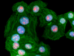
HeLa cells were fixed with 2% PFA for ten minutes, permeabil... 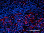
IHC staining using purified anti-HMGB1 (clone 3E8) on formal... -
PE anti-HMGB1
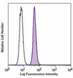
Hela cells were fixed and permeabilized with FOXP3 Fix/Perm ... -
Alexa Fluor® 594 anti-HMGB1
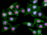
HeLa cells were fixed with 1% paraformaldehyde (PFA) for 10 ... -
Alexa Fluor® 488 anti-HMGB1
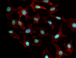
HeLa cells were fixed with 1% paraformaldehyde (PFA) for ten... -
Alexa Fluor® 647 anti-HMGB1
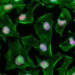
HeLa cells were fixed with 1% paraformaldehyde (PFA) for ten... -
Ultra-LEAF™ Purified anti-HMGB1
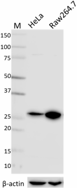
Whole cell lysates (15 µg protein) from HeLa and Raw264.7 ce... -
Direct-Blot™ HRP anti-HMGB1
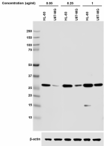
Western blot analysis of 15 µg cell lysates from HL-60 (High... -
Brilliant Violet 421™ anti-HMGB1
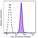
Hela cells were fixed and permeabilized with FOXP3 Fix/Perm ...




Follow Us