- Clone
- O91D11 (See other available formats)
- Regulatory Status
- RUO
- Other Names
- IL-36 alpha, IL-1F6, FIL1 epsilon, Interleukin-1 epsilon, IL-1 epsilon
- Isotype
- Mouse IgG1, κ
- Ave. Rating
- Submit a Review
- Product Citations
- publications

-

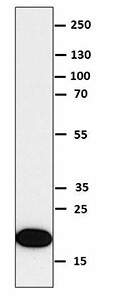
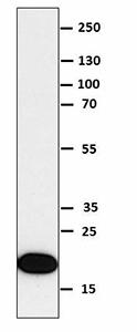
Recombinant human IL-36α (50 ng) was resolved by electrophoresis, transferred to nitrocellulose, and probed with purified monoclonal anti-IL-36α antibody (clone O91D11) antibody. Proteins were visualized using a goat anti-mouse-IgG secondary antibody conjugated to HRP and chemiluminescence detection.
IL-36α is one of the IL-36 cytokines that are part of the IL-1 family. Like other IL-1 family members, IL-36α requires N-terminal processing to gain more potent bioactivity. Currently, the proteases responsible for processing IL-36α are unknown. IL-36α signals through IL-36R/IL-1RAcP, which results in MAPK, Erk1/2, and JNK activation. IL-36α is implicated in skin homeostasis, and it is overexpressed in psoriatic lesional skin. Transgenic mice overexpressing IL-36α in skin have an inflammatory skin condition, which shows some characteristics of human psoriasis, including thickened scaly skin, acanthosis, hyperkeratosis, and dermis infiltration. EGF regulates the expression of IL-36α in the skin. IL-36α can also be detected in adipose tissue. IL-36α reduces adipocyte differentiation and also induces inflammatory gene expression in mature adipocytes. In the lungs, the expression of IL-36α is increased in response to inflammatory stimuli. Intratracheal instillation of recombinant mouse IL-36α induces CXCL1 and CXCL2 expression along with neutrophil influx in the lungs. IL-36α, IL-36β, and IL-36γ induce in vitro expression of the RN of multiple cytokines (IL-6, IL-12 p40, CXCL1, CCL1, IL-12 p35, IL-1β, IL-23 p19, GM-CSF, CXCL10, TNF-α, CCL3, VCAM-1, and ICAM-1) in mouse bone marrow-derived dendritic cells and CD4 T cells obtained from normal mice. IL-36α expression is elevated in chronic kidney disease and in rheumatoid arthritis synovium, and its decreased expression correlates with a poor prognosis in hepatocellular carcinoma.
Product DetailsProduct Details
- Verified Reactivity
- Human
- Antibody Type
- Monoclonal
- Host Species
- Mouse
- Immunogen
- Full length recombinant human IL-36α (NP_055255) produced in HEK 293T cell line.
- Formulation
- Phosphate-buffered solution, pH 7.2, containing 0.09% sodium azide.
- Preparation
- The antibody was purified by affinity chromatography.
- Concentration
- 0.5 mg/ml
- Storage & Handling
- The antibody solution should be stored undiluted between 2°C and 8°C.
- Application
-
WB - Quality tested
- Recommended Usage
-
Each lot of this antibody is quality control tested by Western blotting. For Western blotting, the suggested use of this reagent is 1.0 - 2.5 µg per ml. It is recommended that the reagent be titrated for optimal performance for each application.
- RRID
-
AB_2565642 (BioLegend Cat. No. 676402)
Antigen Details
- Structure
- Full length IL-36α contains 167 amino acids with a predicted molecular weight of 17.7 kD. Proccessing at the N-terminal of IL-36α results in higher potency.
- Distribution
-
Monocytes, T and B cells, spleen, bone marrow, tonsils, lymph nodes, skin, lung, fetal brain, stomach, and adipose tissue.
- Function
- IL-36α participates in inflammatory reactions, induces multiple cytokines in vivo and in vitro, and is involved in skin homeostasis and reduction of adipocyte differentiation. IL-17 and TNF induce IL-36α expression in keratinocytes, and IL-22 synergizes this induction. EGF also regulates the expression of IL-36α in the skin.
- Interaction
- Macrophages, adipocytes, lung fibroblasts, bone marrow dendritic cells, and T cells.
- Ligand/Receptor
- IL36R/IL-RAcP.
- Cell Type
- B cells, Dendritic cells, Monocytes, T cells
- Biology Area
- Angiogenesis, Cell Biology, Immunology, Innate Immunity, Signal Transduction
- Molecular Family
- Cytokines/Chemokines
- Antigen References
-
1. Gresnigt MS and van de Veerdonk FL. 2013. Semin Immunol. 6:458.
2. Towne JE and Sims JE. 2012. Curr. Opin. Pharmacol. 12:486.
3. Vigne S, et al. 2011. Blood 118:5813.
4. Towne JE, et al. 2011. J. Biol. Chem. 286:42594.
5. Chustz RT, et al. 2011. J. Respir. Cell Mol. Biol. 45:145.
6. Vos JB, et al. 2005. Physiol. Genomics 21:324.
7. Pan QZ, et al. 2013. Cancer Immunol. Immunother. 62:1675. - Gene ID
- 27179 View all products for this Gene ID
- UniProt
- View information about IL-36alpha on UniProt.org
Related Pages & Pathways
Pages
Related FAQs
Other Formats
View All IL-36α Reagents Request Custom Conjugation| Description | Clone | Applications |
|---|---|---|
| Purified anti-IL-36α | O91D11 | WB |
Compare Data Across All Formats
This data display is provided for general comparisons between formats.
Your actual data may vary due to variations in samples, target cells, instruments and their settings, staining conditions, and other factors.
If you need assistance with selecting the best format contact our expert technical support team.
-
Purified anti-IL-36α

Recombinant human IL-36α (50 ng) was resolved by electrophor...


Follow Us