- Clone
- M3907B02 (See other available formats)
- Regulatory Status
- RUO
- Other Names
- Macrophage migration inhibitory factor (MMIF), Glycosylation inhibiting factor (GIF), GLIF, Phenylpyruvate Tautomerase, L-Dopachrome Tautomerase, L-Dopachrome Isomerase
- Isotype
- Rat IgG2a, κ
- Ave. Rating
- Submit a Review
- Product Citations
- publications

MIF (human macrophage migration inhibitory factor) is a non-glycosylated polypeptide expressed by multiple cell types, which include activated T cells, macrophages, eosinophils, epithelial cells, and endothelial cells. It is secreted by response to diverse inflammatory stimuli, and has been associated with a clear pro-inflammatory and proatherogenic role in multiple studies involving patients and animals. MIF also positively correlates with obesity as well as insulin resistance and contributes to adipose tissue inflammation by modulating ATM functions. Furthermore, MIF has been described as a protein that plays an essential role in both innate and acquired immunity.
Product DetailsProduct Details
- Verified Reactivity
- Mouse
- Antibody Type
- Monoclonal
- Host Species
- Rat
- Immunogen
- In-house recombinant mouse MIF.
- Formulation
- Phosphate-buffered solution, pH 7.2, containing 0.09% sodium azide.
- Preparation
- The antibody was purified by affinity chromatography.
- Concentration
- 0.5 mg/ml
- Storage & Handling
- The antibody solution should be stored undiluted between 2°C and 8°C.
- Application
-
ELISA Capture - Quality tested
- Recommended Usage
-
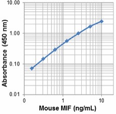
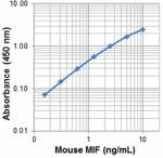
Each lot of this antibody is quality control tested by ELISA assay. For ELISA capture applications, a concentration of 3.0 µg/mL is recommended. To obtain a linear standard curve, serial dilutions of MIF recombinant protein ranging from 10 to 0.16 ng/mL is recommended for each ELISA plate. It is recommended that the reagent be titrated for optimal performance.
- Application Notes
-
ELISA Capture: The purified M3907B02 antibody is useful as the capture antibody in a sandwich ELISA assay, when used in conjunction with the biotinylated Poly5240 antibody (Cat. No. 524004) as the detecting antibody.
Note: For testing mouse MIF in serum, plasma, cell culture supernatant, and cell lysate, BioLegend's LEGEND MAX™ Kits are specially developed and recommended. - RRID
-
AB_2686958 (BioLegend Cat. No. 532502)
Antigen Details
- Structure
- 115 amino acids with a predicted molecular weight of approximately 12.5 kD.
- Distribution
-
Activated T-cells, macrophages, eosinophils, epithelial cells, and endothelial cells.
- Function
- MIF has been associated with a clear pro-inflammatory and proatherogenic role in cardiovascular diseases. MIF also plays important roles in adipose tissue inflammation and innate and acquired immunity.
- Interaction
- Monocytes and macrophages, mesenchymal cells, and B-cells.
- Ligand/Receptor
- CD44/CD74, CXCR4, CXCR2, and CXCR7.
- Cell Type
- T cells, Macrophages, Eosinophils, Epithelial cells, Endothelial cells
- Biology Area
- Angiogenesis, Apoptosis/Tumor Suppressors/Cell Death, Cell Biology, Cell Cycle/DNA Replication, Immunology, Innate Immunity, Signal Transduction
- Molecular Family
- Cytokines/Chemokines
- Antigen References
-
1. Zhou H, et al. 2009. PLoS One. 4:e6087.
2. Potolicchio I, et al. 2003. J. Biol. Chem. 278:30889.
3. Meyer-Siegler K and Iczkowski K. 2005. BMC Cancer 5:73.
4. Babu SN, et al. 2012. Asian Pac. J. Cancer Prev. 13:1737.
5. van der Vorst EP, et al. 2015. Front Immunol. 6:373.
6. Kim BS, et al. 2015. Exp. Mol. Med. 47:e161.
7. Sanchez-Zamora YI, et al. 2014. J. Diabetes Res. 2014:804519.
8. Gilliver SC, et al. 2011. Exp. Dermatol. 20:1.
9. Bach JP, et al. 2008. Oncology 75:127. - Gene ID
- 17319 View all products for this Gene ID
- UniProt
- View information about MIF on UniProt.org
Related Pages & Pathways
Pages
Related FAQs
Other Formats
View All MIF Reagents Request Custom Conjugation| Description | Clone | Applications |
|---|---|---|
| Purified anti-mouse MIF | M3907B02 | ELISA Capture |
Compare Data Across All Formats
This data display is provided for general comparisons between formats.
Your actual data may vary due to variations in samples, target cells, instruments and their settings, staining conditions, and other factors.
If you need assistance with selecting the best format contact our expert technical support team.


Follow Us