


- Clone
- NSE-P1 (See other available formats)
- Regulatory Status
- RUO
- Other Names
- Gamma-enolase, neural enolase, neuron-specific enolase, 2-phospho-D-glycerate hydrolyase, ENO2, NSE
- Previously
-
Covance Catalog# MMS-518P
- Isotype
- Mouse IgG1, κ
- Ave. Rating
- Submit a Review
- Product Citations
- publications

-

IHC staining of purified anti-NSE antibody (clone NSE-P1) on formalin-fixed paraffin-embedded rat brain tissue. Following antigen retrieval using Sodium Citrate H.I.E.R., the tissue was incubated with 5 µg/ml of the primary antibody overnight at 4°C, followed by incubation with Alexa Fluor® 488 goat anti-mouse IgG for two hours at room temperature. The image was captured with a 40X objective. Scale bar: 50 µm -

IHC staining of purified anti-NSE antibody (clone NSE-P1) on formalin-fixed paraffin-embedded mouse brain tissue. Following antigen retrieval using Sodium Citrate H.I.E.R., the tissue was incubated with 10 µg/ml of the primary antibody overnight at 4°C, followed by incubation with Alexa Fluor® 594 goat anti-mouse IgG for two hours at room temperature. Nuclei were counterstained with DAPI. The image was captured with a 40X objective. -

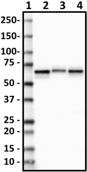
Western blot of purified anti-NSE antibody (clone NSE-P1). Lane 1: Molecular weight marker; Lane 2: 20 µg of human brain lysate. Lane 3: 20 µg of mouse brain lysate. Lane 4: 20 µg of rat brain lysate. The blot was incubated with 0.5 µg/mL of the primary antibody overnight at 4°C, followed by incubation with HRP-labeled goat anti-mouse IgG (Cat. No. 405306). Enhanced chemiluminescence (Cat. No. 426303) was used as the detection system. -

ICC staining of purified anti-NSE antibody (clone NSE-P1) on SH-SY5Y Cells. The cells were fixed with 4% PFA, permeabilized with a buffer containing 0.1% Triton X-100 and 0.25% BSA, and blocked with 2% normal goat serum and 0.02% BSA. The cells were then incubated with 0.5 µg/mL of the primary antibody overnight at 4°C, followed by incubation with 2.5 µg/mL of Alexa Fluor® 594 goat anti-mouse IgG for one hour at room temperature. The cells were co-stained with Flash Phalloidin™ Green 488 (Cat. No. 424201). The slide was mounted with fluoromount G with DAPI. The image was captured with a 60X objective. Scale bar: 20 µm
| Cat # | Size | Price | Save |
|---|---|---|---|
| 804908 | 25 µL | ¥24,640 | |
| 804901 | 200 µL | ¥60,720 |
Mammalian enolase is composed of 3 isozyme subunits, alpha, beta and gamma, which can form homodimers or heterodimers. Expression of these isozymes can be cell-type and development-specific. This gene encodes gamma enolase (enolase 2) and is expressed in mature neurons and cells of neuronal origin. The alpha/alpha homodimers are expressed in embryo and in most adult tissues. The alpha/beta heterodimers and the beta/beta homodimers are found in striated muscle, and the alpha/gamma heterodimers and the gamma/gamma homodimers are expressed in neurons. Neuron specific enolase is found in elevated concentrations in plasma and certain neoplasias. These include pediatric neuroblastoma and small cell lung cancer.
Product DetailsProduct Details
- Verified Reactivity
- Human, Mouse, Rat
- Antibody Type
- Monoclonal
- Host Species
- Mouse
- Immunogen
- This antibody was raised against a synthetic peptide corresponding to amino acids 416-433. It recognizes the sequence LGDEARFAGHNFRNPSVL.
- Formulation
- Phosphate-buffered solution + 0.03% thimerosal.
- Preparation
- The antibody was purified by affinity chromatography.
- Concentration
- 1 mg/mL
- Storage & Handling
- The antibody solution should be stored undiluted between 2°C and 8°C. Please note the storage condition for this antibody has been changed from -20°C to between 2°C and 8°C. You can also check your vial or your CoA to find the most accurate storage condition for this antibody.
- Application
-
IHC-P - Quality tested
WB, ICC - Verified
ELISA - Reported in the literature, not verified in house - Recommended Usage
-
Each lot of this antibody is quality control tested by formalin-fixed paraffin-embedded immunohistochemical staining. For immunohistochemistry, a concentration range of 5.0 - 10.0 µg/mL is suggested. For Western blotting, the suggested use of this reagent is 0.5 - 5.0 µg per mL. For immunocytochemistry, a concentration range of 0.5 - 10 μg/mL is recommended. It is recommended that the reagent be titrated for optimal performance for each application.
- Application Notes
-
The reactivity of Alexa Fluor® 647 and Alexa Fluor® 488 anti-NSE antibody was verified in FFPE mouse and rat tissue.
Additional reported applications (for the relevant formats) include: ELISA2 and immunohistochemistry1,3,4. -
Application References
(PubMed link indicates BioLegend citation) -
- Yoo J, et al. 2002. Arch. Pathol. Lab Med. 126(8):979. (IHC-P)
- Duncan ME, et al. 1992. J. Immuno. Methods. 151:227 (ELISA)
- Chetty R. 2000. Pathology. 32(3):209. (IHC-P)
- Murray GI, et al. 1993. J. Clin. Pathol. 46:993. (IHC-P)
- Product Citations
-
- RRID
-
AB_2728530 (BioLegend Cat. No. 804908)
AB_2564673 (BioLegend Cat. No. 804901)
Antigen Details
- Structure
- Gamma enolase is a 434 amino acid protein with a molecular mass of 47 kD.
- Distribution
-
Tissue distribution: Nervous and musculoskeletal systems.
Cellular distribution: Secreted, cytosol, plasma membrane, cytoskeleton, and nucleus. - Function
- Gamma enolase has neurotrophic and neuroprotective properties on a broad spectrum of central nervous system (CNS) neurons. It binds, in a calcium-dependent manner, to cultured neocortical neurons and promotes cell survival.
- Cell Type
- Mature Neurons, Neural Stem Cells
- Biology Area
- Cell Biology, Neuroscience, Neuroscience Cell Markers, Stem Cells
- Molecular Family
- Phospho-Proteins
- Antigen References
-
- Muoio B, et al. 2017. Int J Biol Markers. 10.5301/ijbm.5000286.
- Huang L, et al. 2017. Oncotarget. 8(38): 64358.
- Isgro MA, et al. 2015. Adv Exp Med Biol. 867:125.
- Zaheer S, et al. 2013. Ann Indian Acad Neurol. 16(4):504.
- Gene ID
- 2026 View all products for this Gene ID
- UniProt
- View information about NSE on UniProt.org

Related FAQs
Other Formats
View All NSE Reagents Request Custom Conjugation| Description | Clone | Applications |
|---|---|---|
| Purified anti-NSE | NSE-P1 | IHC-P,WB,ICC,ELISA |
| Alexa Fluor® 594 anti-NSE | NSE-P1 | IHC-P,ICC |
| Alexa Fluor® 488 anti-NSE | NSE-P1 | IHC-P |
| Alexa Fluor® 647 anti-NSE | NSE-P1 | IHC-P |
| Spark YG™ 570 anti-NSE | NSE-P1 | IHC-P |
Customers Also Purchased
Compare Data Across All Formats
This data display is provided for general comparisons between formats.
Your actual data may vary due to variations in samples, target cells, instruments and their settings, staining conditions, and other factors.
If you need assistance with selecting the best format contact our expert technical support team.
-
Purified anti-NSE

Western blot of purified anti-NSE antibody (clone NSE-P1). L... 
IHC staining of purified anti-NSE antibody (clone NSE-P1) on... 
IHC staining of purified anti-NSE antibody (clone NSE-P1) on... 
ICC staining of purified anti-NSE antibody (clone NSE-P1) on... -
Alexa Fluor® 594 anti-NSE

ICC staining of Alexa Fluor® 594 anti-NSE antibody (clone NS... 
IHC staining of Alexa Fluor® 594 anti-NSE antibody (clone NS... -
Alexa Fluor® 488 anti-NSE

IHC staining of Alexa Fluor® 488 anti-NSE antibody (clone NS... 
IHC staining of Alexa Fluor® 488 anti-NSE antibody (clone NS... -
Alexa Fluor® 647 anti-NSE

IHC staining of Alexa Fluor® 647 anti-NSE antibody (clone NS... 
IHC staining of Alexa Fluor® 647 anti-NSE Antibody (clone NS... -
Spark YG™ 570 anti-NSE

IHC staining of Spark YG™ 570 anti-NSE antibody (clone NSE-P... 
IHC staining of Spark YG™ 570 anti-NSE antibody (clone NSE-P... 
IHC staining of Spark YG™ 570 anti-NSE antibody (clone NSE-P...


Follow Us