


- Clone
- A15165D (See other available formats)
- Regulatory Status
- RUO
- Other Names
- PARK2, AR-JP, LPRS2, PDJ, PRKN, E3 ubiquitin-protein ligase parkin, Parkin 2, E3 ubiquitin ligase, parkinson disease protein 2, parkinson juvenile disease protein 2, Parkinson disease (autosomal recessive, juvenile) 2
- Isotype
- Mouse IgG2b, κ
- Ave. Rating
- Submit a Review
- Product Citations
- publications

-

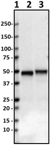
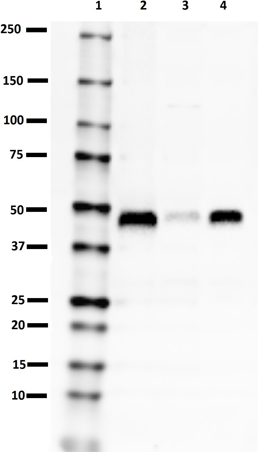
Western blot of purified anti-Parkin antibody (clone A15165D). Lane 1: Molecular weight marker; Lane 2: 20 µg of human brain lysate. The blot was incubated with 2 µg/mL of the primary antibody overnight at 4°C, followed by incubation with HRP goat anti-mouse IgG antibody (Cat. No. 405306). Enhanced chemiluminescence was used as the detection system. -

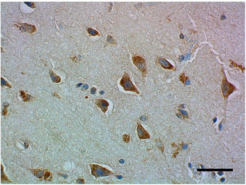
IHC staining of purified anti-Parkin antibody (clone A15165D) on formalin-fixed paraffin-embedded human brain tissue. Following antigen retrieval using Sodium Citrate H.I.E.R., the tissue was incubated with 10 µg/mL of the primary antibody overnight at 4°C. BioLegend’s Ultra Streptavidin (USA) HRP Detection Kit (Multi-Species, DAB, Cat. No. 929901) was used for detection followed by hematoxylin counterstaining, according to the protocol provided. The image was captured with a 40X objective. Scale bar: 50 µm -

IHC staining of purified anti-Parkin antibody (clone A15165D) on formalin-fixed paraffin-embedded Parkinson’s disease substantia nigra tissue. Following antigen retrieval using Sodium Citrate H.I.E.R., the tissue was incubated with 10 µg/mL of the primary antibody overnight at 4°C. BioLegend’s Ultra Streptavidin (USA) HRP Detection Kit (Multi-Species, DAB, Cat. No. 929901) was used for detection followed by hematoxylin counterstaining, according to the protocol provided. The image was captured with a 40X objective. Scale bar: 50 µm -

IHC staining of purified anti-Parkin antibody (clone A15165D) on formalin-fixed paraffin-embedded human brain tissue. Following antigen retrieval using Sodium Citrate H.I.E.R., the tissue was incubated with 10 µg/mL of the primary antibody overnight at 4°C. BioLegend’s Ultra Streptavidin (USA) HRP Detection Kit (Multi-Species, Cat. No. 929701) and Boost DAB Enhancer (Cat. No. 926701) were used for detection followed by hematoxylin counterstaining, according to the protocol provided. The image was captured with a 40X objective. Scale bar: 50 µm -

IHC staining of purified anti-Parkin antibody (clone A15165D) on formalin-fixed paraffin-embedded Parkinson’s disease substantia nigra tissue. Following antigen retrieval using Sodium Citrate H.I.E.R., the tissue was incubated with 10 µg/mL of the primary antibody overnight at 4°C. BioLegend’s Ultra Streptavidin (USA) HRP Detection Kit (Multi-Species, Cat. No. 929701) and Boost DAB Enhancer (Cat. No. 926701) were used for detection followed by hematoxylin counterstaining, according to the protocol provided. The image was captured with a 40X objective. Scale bar: 50 µm
| Cat # | Size | Price | Save |
|---|---|---|---|
| 870501 | 25 µg | ¥23,320 | |
| 870502 | 100 µg | ¥60,720 |
Parkin (also known as PARK2, AR-JP, PRKN) is a protein encoded by the PARK2 gene in humans. It is a component of the E3 ubiquitin ligase complex that mediates the targeting of proteins for degradation. Mutations in the PARK2 gene cause a familial form of Parkinson’s disease called autosomal recessive juvenile parkinsonism (AR-JP).
Product DetailsProduct Details
- Verified Reactivity
- Human
- Antibody Type
- Monoclonal
- Host Species
- Mouse
- Immunogen
- Recombinant full-length human Parkin protein
- Formulation
- Phosphate-buffered solution, pH 7.2, containing 0.09% sodium azide
- Preparation
- The antibody was purified by affinity chromatography.
- Concentration
- 0.5 mg/mL
- Storage & Handling
- The antibody solution should be stored undiluted between 2°C and 8°C.
- Application
-
WB - Quality tested
IHC-P - Verified - Recommended Usage
-
Each lot of this antibody is quality control tested by western blotting. For western blotting, the suggested use of this reagent is 2.0 - 10 µg/mL. For immunohistochemistry on formalin-fixed paraffin-embedded tissue sections, a concentration of 10 µg/mL is suggested. It is recommended that the reagent be titrated for optimal performance for each application.
- Product Citations
-
- RRID
-
AB_2820194 (BioLegend Cat. No. 870501)
AB_2820194 (BioLegend Cat. No. 870502)
Antigen Details
- Structure
- Human parkin is a 465 amino acid protein with a predicted molecular mass of ~52 kD.
- Distribution
-
Tissue distribution: Brain, heart, testis, and skeletal muscle
Cellular distribution: Cytosol, mitochondria, nucleus, and endoplasmic reticulum - Function
- Parkin is involved in protein ubiquitination, protein degradation, mitochondria function, and neurodegeneration.
- Interaction
- Ubiquitin, PINK1
- Ligand/Receptor
- BCL2, SYT11, CCNE1, GPR37, RHOT1/MIRO1, MFN1
- Cell Type
- Neurons
- Biology Area
- Cell Biology, Mitochondrial Function, Neurodegeneration, Neuroinflammation, Neuroscience, Neuroscience Cell Markers, Protein Misfolding and Aggregation, Protein Trafficking and Clearance, Ubiquitin/Protein Degradation
- Molecular Family
- Ligases
- Antigen References
-
- Tay SP, et al. 2010. J Biol Chem. 285:29231-8.
- Pawlyk AC, et al. 2003. J Biol Chem. 278:48120-8
- Gene ID
- 5071 View all products for this Gene ID
- UniProt
- View information about Parkin on UniProt.org

Related FAQs
Other Formats
View All Parkin Reagents Request Custom Conjugation| Description | Clone | Applications |
|---|---|---|
| Purified anti-Parkin | A15165D | WB,IHC-P |
Customers Also Purchased
Compare Data Across All Formats
This data display is provided for general comparisons between formats.
Your actual data may vary due to variations in samples, target cells, instruments and their settings, staining conditions, and other factors.
If you need assistance with selecting the best format contact our expert technical support team.
-
Purified anti-Parkin

Western blot of purified anti-Parkin antibody (clone A15165D... 
IHC staining of purified anti-Parkin antibody (clone A15165D... 
IHC staining of purified anti-Parkin antibody (clone A15165D... 
IHC staining of purified anti-Parkin antibody (clone A15165D... 
IHC staining of purified anti-Parkin antibody (clone A15165D...


Follow Us