- Regulatory Status
- RUO
- Other Names
- Interleukin-36 beta, Interleukin-1 family member 8, IL-1F8, Interleukin-1 homolog 2, IL-1H2
- Ave. Rating
- Submit a Review
- Product Citations
- publications

-

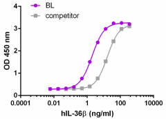
Dose-dependent stimulation of IL-8 secretion in A431 cells.
| Cat # | Size | Price | Save |
|---|---|---|---|
| 761102 | 10 µg | ¥44,380 | |
| 761104 | 25 µg | ¥87,560 |
The IL-1 family is a group of cytokines comprised of 11 members, including the IL-36 cytokines, IL-36α, β and γ (previously known as IL-1F6, IL-1F8, and IL-1F9). Like other IL-36 cytokines, IL-36β signals through IL-1Rrp2 and IL-1RAcP and activate NF-κB and MAPKs. IL-36β functions as a chemokine and induces migration of bone marrow-derived dendritic cells (BMDCs) and CD4+ T cells. IL-36β induces proinflammatory cytokines, including IL-12, IL-1β, IL-6, and TNF-α in BMDCs. Furthermore, it has been shown that IL-36β synergizes with IL-12 to enhance Th1 polarization. IL-36β is also expressed in neurons and glia cells, but recombinant IL-36β does not induce the classical IL-1 response in brain cells. In a cultured keratinocyte system, IL-36β can induce robust expression of many chemokines in macrophages, T cells, and neutrophils. The secretion of antimicrobial peptides, including HBD-2, HBD-3, lipocalin 2, CAMP, elafin, serpinB1, and IL-8 can be induced by IL-36β in keratinocytes in culture. IL-36β, can also upregulate MMP9 and MMP19 mRNA in cultured keratinocytes. IL-36β has been implicated in the development of human psoriasis. IL-36β is expressed in the skin and the expression can be upregulated by IL-1β, TNF-α, flagellin, and poly (I:C). The expression of IL-36β is also increased in plaque psoriasis. In addition, IL-36β can be detected in mononuclear cells that infiltrate lesional skin. In mouse models of psoriasis, IL-36β is significantly upregulated in inflamed skin.
Product DetailsProduct Details
- Source
- Human IL-36β, amino acids Arg5-Glu157 (Accession# NP_775270), was expressed in E. coli.
- Molecular Mass
- The 153 amino acid recombinant protein has a predicted molecular mass of approximately 17 kD. The DTT-reduced and non-reduced protein migrates at approximately 17 kD by SDS-PAGE. The predicted N-terminal amino acid is Arg.
- Purity
- >95% as determined by Coomassie stained SDS-PAGE under reducing conditions.
- Formulation
- 0.22 µm filtered protein solution is in 1x PBS.
- Endotoxin Level
- Less than 0.01 ng per µg cytokine as determined by the LAL method.
- Concentration
- 10 and 25 µg sizes are bottled at 200 µg/mL.
- Storage & Handling
- Unopened vial can be stored between 2°C and 8°C for up to 2 weeks, at -20°C for up to six months, or at -70°C or colder until the expiration date. For maximum results, quick spin vial prior to opening. The protein can be aliquoted and stored at -20°C or colder. Stock solutions can also be prepared at 50 - 100 µg/mL in appropriate sterile buffer, carrier protein such as 0.2 - 1% BSA or HSA can be added when preparing the stock solution. Aliquots can be stored between 2°C and 8°C for up to one week and stored at -20°C or colder for up to 3 months. Avoid repeated freeze/thaw cycles.
- Activity
- The ED50 is 0.8-4.8 ng/mL as determined by a dose-dependent stimulation of IL-8 secretion in A431 cells.
- Recommended Usage
-
Bioassay
- Application Notes
-
BioLegend carrier-free recombinant proteins provided in liquid format are shipped on blue-ice. Our comparison testing data indicates that when handled and stored as recommended, the liquid format has equal or better stability and shelf-life compared to commercially available lyophilized proteins after reconstitution. Our liquid proteins are verified in-house to maintain activity after shipping on blue ice and are backed by our 100% satisfaction guarantee. If you have any concerns, contact us at tech@biolegend.com.
Antigen Details
- Structure
- IL-1 family cytokine.
- Distribution
-
Skin, tonsils, bone marrow, heart, placenta, lung, testis, monocytes, B cells, neurons, and glia cells.
- Function
- IL-36β induces proinflammatory cytokines, synergizes with IL-12 to promote Th1 polarization, and induces antimicrobial peptides. IL-36β is upregulated by IL-1β, TNF-α, flagellin, and poly (I:C); its activity is significantly increased by N-terminal processing.
- Interaction
- Keratinocytes, dendritic cells, macrophages, CD4+ T cells.
- Ligand/Receptor
- Heterodimeric receptor IL-1Rrp2 and IL-1RAcP.
- Biology Area
- Angiogenesis, Cell Biology, Immunology, Innate Immunity, Signal Transduction
- Molecular Family
- Cytokines/Chemokines
- Antigen References
-
1. Towne JE, et al. 2004. J. Biol. Chem. 279:13677.
2. Barksby HE, et al. 2007. Clin. Exp. Immunol. 149:217.
3. Towne JE, et al. 2011. J. Biol. Chem. 286:42594.
4. Wang P, et al. 2005. Cytokine. 29:245.
5. Gresnigt MS, van de Veerdonk FL. 2013. Semin. Immunol. 25:458.
6. Tortola L, et al. 2012. J. Clin. Invest. 122:3965.
7. Johnston A, et al. 2011. J. Immunol. 186:2613. - Gene ID
- 27177 View all products for this Gene ID
- UniProt
- View information about IL-36beta on UniProt.org
Related Pages & Pathways
Pages
Related FAQs
- Why choose BioLegend recombinant proteins?
-
• Each lot of product is quality-tested for bioactivity as indicated on the data sheet.
• Greater than 95% Purity or higher, tested on every lot of product.
• 100% Satisfaction Guarantee for quality performance, stability, and consistency.
• Ready-to-use liquid format saves time and reduces challenges associated with reconstitution.
• Bulk and customization available. Contact us.
• Learn more about our Recombinant Proteins. - How does the activity of your recombinant proteins compare to competitors?
-
We quality control each and every lot of recombinant protein. Not only do we check its bioactivity, but we also compare it against other commercially available recombinant proteins. We make sure each recombinant protein’s activity is at least as good as or better than the competition’s. In order to provide you with the best possible product, we ensure that our testing process is rigorous and thorough. If you’re curious and eager to make the switch to BioLegend recombinants, contact your sales representative today!
- What is the specific activity or ED50 of my recombinant protein?
-
The specific activity range of the protein is indicated on the product datasheets. Because the exact activity values on a per unit basis can largely fluctuate depending on a number of factors, including the nature of the assay, cell density, age of cells/passage number, culture media used, and end user technique, the specific activity is best defined as a range and we guarantee the specific activity of all our lots will be within the range indicated on the datasheet. Please note this only applies to recombinants labeled for use in bioassays. ELISA standard recombinant proteins are not recommended for bioassay usage as they are not tested for these applications.
- Have your recombinants been tested for stability?
-
Our testing shows that the recombinant proteins are able to withstand room temperature for a week without losing activity. In addition the recombinant proteins were also found to withstand four cycles of freeze and thaw without losing activity.
- Does specific activity of a recombinant protein vary between lots?
-
Specific activity will vary for each lot and for the type of experiment that is done to validate it, but all passed lots will have activity within the established ED50 range for the product and we guarantee that our products will have lot-to-lot consistency. Please conduct an experiment-specific validation to find the optimal ED50 for your system.
- How do you convert activity as an ED50 in ng/ml to a specific activity in Units/mg?
-
Use formula Specific activity (Units/mg) = 10^6/ ED50 (ng/mL)


Follow Us