- Clone
- A15105B (See other available formats)
- Regulatory Status
- RUO
- Other Names
- Calprotectin, Human S100A8 (Myeloid-related protein, MRP 8), S100A9 (MRP14), calgranulin A and B, L1, cystic fibrosis antigen, and 27E10 antigen, P8, MIF, NIF, CAGA, CFAG, CGLA, L1Ag, MRP8, CP-10, MA387, 60B8AG
- Isotype
- Mouse IgG2b, κ
- Ave. Rating
- Submit a Review
- Product Citations
- publications

-

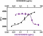
Recombinant human S100A8/A9 heterodimer stimulates proliferation of astrocytes in a dose-dependent manner (black circles). Proliferation induced by human S100A8/A9 heterodimer is neutralized (purple circles) by increasing concentrations of anti-human S100A8/A9 monoclonal antibody (Clone A15015B). The ND50 is typically 2 - 8 µg/mL.
| Cat # | Size | Price | Save |
|---|---|---|---|
| 697503 | 100 µg | ¥44,820 | |
| 697504 | 1 mg | ¥118,690 |
S100A8/A9, also known as Calprotectin, is a heterodimeric protein that belongs to the family of low molecular weight, calcium binding S100 proteins with two EF-hand calcium binding domains. Calcium and zinc binding influences the S100A8/A9 conformation and oligomerization state, which includes the self-assembly into homo- and heterodimers and larger oligomers. S100A8/A9 is elevated in blood and other body fluids in many pathological conditions including rheumatoid arthritis, inflammatory bowel diseases, viral or microbial infections, and tumors. S100A8/A9 is identified as an endogenous activator of TLR4 and is involved in promoting the inflammatory response in infections. Neutrophil-derived S100A8/A9 inhibits growth of Staphylococcus aureus through the chelation of manganese and zinc. S100A8/A9 is also identified as a potent amplifier of inflammation in autoimmunity as well as in cancer development and tumor spread. Injury induced by S100A8/A9 stimulates IP-10 production in monocytes and macrophages. In addition, the S100A8/A9 heterodimer also promotes astrocyte proliferation.
Product DetailsProduct Details
- Verified Reactivity
- Human
- Antibody Type
- Monoclonal
- Host Species
- Mouse
- Immunogen
- Recombinant human A100A8/A9
- Formulation
- 0.2 µm filtered in phosphate-buffered solution, pH 7.2, containing no preservative.
- Endotoxin Level
- Less than 0.01 EU/µg of the protein (< 0.001 ng/µg of the protein) as determined by the LAL test.
- Preparation
- The Ultra-LEAF™ (Low Endotoxin, Azide-Free) antibody was purified by affinity chromatography.
- Concentration
- The antibody is bottled at the concentration indicated on the vial, typically between 2 mg/mL and 3 mg/mL. Older lots may have also been bottled at 1 mg/mL. To obtain lot-specific concentration and expiration, please enter the lot number in our Certificate of Analysis online tool.
- Storage & Handling
- The antibody solution should be stored undiluted between 2°C and 8°C. This Ultra-LEAF™ solution contains no preservative; handle under aseptic conditions.
- Application
-
Block - Quality tested
- Recommended Usage
-
Each lot of this antibody is quality control tested by blocking activity. The ND50 is 2 - 8 µg/mL. It is recommended that the reagent be titrated for optimal performance for each application.
- RRID
-
AB_2687183 (BioLegend Cat. No. 697503)
AB_2687183 (BioLegend Cat. No. 697504)
Antigen Details
- Structure
- Non-covalent heterodimer.
- Distribution
-
Neutrophils, monocytes, macrophages, gingival keratinocytes, granulocytes, osteoclasts, fibroblasts, and microvascular endothelial cells.
- Function
- Pro-inflammatory molecule that possesses antimicrobial properties. Is up-regulated in neutrophils and monocytes at the sites of inflammation.
- Interaction
- Astrocytes, neutrophils, and monocytes.
- Ligand/Receptor
- RAGE, TLR4
- Cell Type
- Neutrophils, Monocytes, Macrophages, Granulocytes, Osteoclasts, Fibroblasts, Endothelial cells
- Biology Area
- Cell Biology, Neuroscience, Stem Cells, Synaptic Biology
- Molecular Family
- Growth Factors, Cytokines/Chemokines
- Antigen References
-
1. Sampson B, et al. 2002. Lancet. 360:1742-5.
2. Vogl T, et al. 2007. Nat. Med. 13:1042-9.
3. Semprini S, et al. 2002. Hum. Genet. 111:310-3.
4. Corbin BD, et al. 2008. Science 319:962-5.
5. Gopal R, et al. 2013. Am. J. Respir. Crit. Care Med. 188:1137-46.
6. Siegenthaler G, et al. 1997. J. Biol. Chem. 14:9371-7.
7. Ryckman C, et al. 2003. J. Immunol. 170:3233-42.
8. Ryu MJ, et al. 2012. J. Biol. Chem. 287:22948-58.
9. Ehrchen JM, et al. 2009. J. Leuko. Biol. 3:557-66.
10. Wang J, et al. 2015. FASEB J. 29(1):250-62.
11. Champaiboon C, et al. 2009. J. Biol. Chem. 284:7078. - Gene ID
- 6279 View all products for this Gene ID
- UniProt
- View information about S100A8/A9 Heterodimer on UniProt.org
Related Pages & Pathways
Pages
Related FAQs
- Do you guarantee that your antibodies are totally pathogen free?
-
BioLegend does not test for pathogens in-house aside from the GoInVivo™ product line. However, upon request, this can be tested on a custom basis with an outside, independent laboratory.
- Does BioLegend test each Ultra-LEAF™ antibody by functional assay?
-
No, BioLegend does not test Ultra-LEAF™ antibodies by functional assays unless otherwise indicated. Due to the possible complexities and variations of uses of biofunctional antibodies in different assays and because of the large product portfolio, BioLegend does not currently perform functional assays as a routine QC for the antibodies. However, we do provide references in which the antibodies were used for functional assays and we do perform QC to verify the specificity and quality of the antibody based on our strict specification criteria.
- Does BioLegend test each Ultra-LEAF™ antibody for potential pathogens?
-
No, BioLegend does not test for pathogens in-house unless otherwise indicated. However, we can recommend an outside vendor to perform this testing as needed.
- Have you tested this Ultra-LEAF™ antibody for in vivo or in vitro applications?
-
We don't test our antibodies for in vivo or in vitro applications unless otherwise indicated. Depending on the product, the TDS may describe literature supporting usage of a particular product for bioassay. It may be best to further consult the literature to find clone specific information.
Other Formats
View All S100A8/A9 Reagents Request Custom Conjugation| Description | Clone | Applications |
|---|---|---|
| Ultra-LEAF™ Purified anti-human S100A8/A9 Heterodimer | A15105B | Block |
Customers Also Purchased
Compare Data Across All Formats
This data display is provided for general comparisons between formats.
Your actual data may vary due to variations in samples, target cells, instruments and their settings, staining conditions, and other factors.
If you need assistance with selecting the best format contact our expert technical support team.
-
Ultra-LEAF™ Purified anti-human S100A8/A9 Heterodimer
Recombinant human S100A8/A9 heterodimer stimulates prolifera...




Follow Us