


- Clone
- A16040D (See other available formats)
- Regulatory Status
- RUO
- Other Names
- Microtubule associated protein tau
- Isotype
- Rat IgG2b, κ
- Ave. Rating
- Submit a Review
- Product Citations
- publications

-

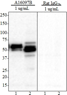
Western blots of anti-Tau, 269-281 antibody (clone A16040D) and isotype-matched control rat IgG2b, κ. Lane 1: 20 µg of normal human brain lysate; Lanes 2 & 3: 20 µg of mouse brain lysates. Blots were incubated overnight at 4°C with 1 or 5 µg/ml of primary antibody, followed by incubation with horseradish peroxidase labeled goat anti-rat IgG secondary antibody. -

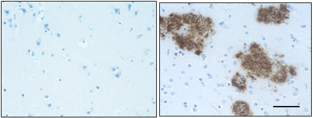
IHC staining of anti-Tau, 269-281 antibody (clone A16040D) and isotype-matched control rat IgG2b on formalin-fixed paraffin-embedded Alzheimer’s disease brain tissue. Following antigen retrieval using Sodium Citrate and 0.25% triton X-100, the tissue was incubated with the primary antibody at 5 µg/ml overnight at 4°C. Biotinylated anti-rat IgG, HRP Streptavidin, and DAB (3,3'-diaminobenzidine) substrate were used as the detection system. -

Direct ELISA of purified anti-Tau, 269-281 antibody (clone A16040D) binding to plate-immobilized human 2N3R and 2N4R Tau proteins. ELISA was performed by coating wells with 100 ng of each recombinant Tau protein. The wells were then incubated with the primary antibody at 37°C for 45 minutes, followed by incubation with horseradish peroxidase labeled goat anti-rat secondary antibody. TMB (3, 3', 5, 5' tetramethylbenzidine) was used as the detection system.
| Cat # | Size | Price | Save |
|---|---|---|---|
| 850601 | 25 µg | ¥24,640 | |
| 850602 | 100 µg | ¥60,720 |
Tau protein promotes microtubule assembly and stability. Tau is abundant in neurons of the central nervous system, and is expressed at low levels in astrocytes and oligodendrocytes. Abnormal hyper-phosphorylation, aggregation, and toxic gain of function of tau is associated with several neurological disorders, including Alzheimer’s disease (AD). The major building block of neurofibrillary lesions in AD brains consists of paired helical filaments (PHFs) of abnormally hyperphosphorylated tau. Six isoforms of tau are generated by alternative splicing of the MAPT gene. These isoforms are distinguished by the number of tubulin binding domains, 3 (3R) or 4 (4R), in the C-terminal of the protein and by one (1N), two (2N), or no (0N) inserts in the N-terminal domain. Tau isoforms are differentially expressed during development.
Product DetailsProduct Details
- Verified Reactivity
- Human, Mouse
- Antibody Type
- Monoclonal
- Host Species
- Rat
- Immunogen
- Tau peptide encompassing amino acid residues 269-281 conjugated to KLH
- Formulation
- Phosphate-buffered solution, pH 7.2, containing 0.09% sodium azide.
- Preparation
- The antibody was purified by affinity chromatography.
- Concentration
- 0.5 mg/ml
- Storage & Handling
- The antibody solution should be stored undiluted between 2°C and 8°C.
- Application
-
WB - Quality tested
IHC-P, ELISA - Verified - Recommended Usage
-
Each lot of this antibody is quality control tested by Western blotting. For Western blotting, the suggested use of this reagent is 1.0 - 5.0 µg per ml. For Immunohistochemistry of paraffin-embedded tissues, the suggested use of this reagent is 1.0 - 5.0 µg per ml. For ELISA, the suggested use of this reagent is 0.02 - 1.0 µg per ml. It is recommended that the reagent be titrated for optimal performance for each application.
- Application Notes
-
This antibody cross-reacts with 2N3R and 2N4R Tau isoforms, and recognizes endogenous human and murine Tau in brain lysates by WB.
- RRID
-
AB_2716012 (BioLegend Cat. No. 850601)
AB_2716012 (BioLegend Cat. No. 850602)
Antigen Details
- Structure
- Unmodified Tau isoforms have an apparent molecular weight ranging from 33-79 kD. Additional high and low molecular weight Tau species have been observed in brain tissues.
- Distribution
-
Tissue distribution: Central nervous system, peripheral ganglia and nerves, kidney, skeletal, and heart muscle.
Cellular distribution: cytoskeleton, nucleus, plasma membrane, and cytosol. - Function
- Tau promotes microtubule assembly and stability. The short tau isoforms allow plasticity of the cytoskeleton whereas the longer isoforms may preferentially play a role in its stabilization.
- Interaction
- Tau interacts with Sequestosome-1, Peptidyl-prolyl cis-trans isomerase FKBP4, Casein kinase I isoform delta, Serine/threonine-protein kinase Sgk1, Laforin, Alpha-synuclein
- Biology Area
- Cell Biology, Neurodegeneration, Neuroscience, Protein Misfolding and Aggregation
- Molecular Family
- Tau
- Antigen References
- Gene ID
- 4137 View all products for this Gene ID
- UniProt
- View information about Tau 269-281 on UniProt.org

Other Formats
View All Tau, 269-281 Reagents Request Custom Conjugation| Description | Clone | Applications |
|---|---|---|
| Purified anti-Tau, 269-281 | A16040D | WB,IHC-P,ELISA |
| Alexa Fluor® 647 anti-Tau, 269-281 | A16040D | IHC-P |
Customers Also Purchased
Compare Data Across All Formats
This data display is provided for general comparisons between formats.
Your actual data may vary due to variations in samples, target cells, instruments and their settings, staining conditions, and other factors.
If you need assistance with selecting the best format contact our expert technical support team.
-
Purified anti-Tau, 269-281

Western blots of anti-Tau, 269-281 antibody (clone A16040D) ... 
IHC staining of anti-Tau, 269-281 antibody (clone A16040D) a... 
Direct ELISA of purified anti-Tau, 269-281 antibody (clone A... -
Alexa Fluor® 647 anti-Tau, 269-281

IHC staining of Alexa Fluor® 647 anti-Tau, 269-281 antibody ...


Follow Us