- Clone
- A17169H (See other available formats)
- Regulatory Status
- RUO
- Other Names
- Platelet Derived Growth Factor Receptor Beta, Beta Platelet-Derived Growth Factor Receptor, PDGFR-B, PDGFR β, CD140B, PDGFR1, PDGF-R-B
- Isotype
- Mouse IgG2b, κ
- Ave. Rating
- Submit a Review
- Product Citations
- publications

-

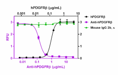
Recombinant human PDGFRβ (Black Circles, Cat. No. 767206) binds to immobilized recombinant human PDGF-BB (Cat. No. 577306) Anti-human PDGFRβ antibody (Purple Squares, Clone A17169H) inhibits the binding in a dose-dependent manner whereas the isotype control (Green Triangles, Cat. No. 400366) does not have this effect. This antibody blocks the binding of 0.3 µg/mL recombinant human PDGFRβ protein to 1 µg/mL of immobilized recombinant human PDGF-BB with an ND50 = 0.08 - 0.6 µg/mL.
| Cat # | Size | Price | Quantity Check Availability | Save | ||
|---|---|---|---|---|---|---|
| 622803 | 100 µg | 216€ | ||||
| 622804 | 1 mg | 572€ | ||||
Select size of product is eligible for a 40% discount! Promotion valid until December 31, 2024. Exclusions apply. To view full promotion terms and conditions or to contact your local BioLegend representative to receive a quote, visit our webpage.
The PDGF family includes five members, PDGF-AA, PDGF-BB, PDGF-AB, PDGF-CC, and PDGF-DD. Two PDGF receptors (α and β) have been described; they belong to the receptor-tyrosine kinase family, Type III group, which also includes c-KIT, FLT3, and M-CSFR. The two PDGF receptor subtypes show different affinities for the dimeric PDGF isoforms. PDGFRα binds to all ligands except PDGF-D, while PDGFRβ binds to PDGF-B and -D only. The two PDGF receptors contain five extracellular immunoglobulin-like domains, a single transmembrane domain, and an intracellular split kinase domain, which is formed by two lobes connected by a flexible polypeptide linker, the kinase insert. The binding of PDGFs to their receptors induces receptor dimerization. PDGF receptors are degraded after activation by a mechanism that involves ligand-induced endocytosis and degradation in lysosomes. The soluble form of PDGFRα was detected in conditioned media from human cell lines. The soluble receptor was derived by proteolytic clipping of the full length PDGFR. Soluble PDGFRβ inhibits hepatic stellate cell activation and attenuates liver fibrosis. Also, soluble PDGFRβ inhibits intraosseous growth of breast cancer cells in nude mice. PDGFRβ mediates recruitment of pericytes and vascular smooth muscle cells to newly formed vessels in response to PDGF-BB secreted by endothelial cells. PDGFRβ knockout embryos develop abnormal glomeruli in the kidney (developmental defect of mesangial cells) capillary microaneurysm, cardiac and placental defects, and die at E16–E19 from hemorrhage. High levels of PDGFRβ expression have been associated with tumor recurrence in primary colorectal cancer, and it correlates with poor disease-free survival in pancreatic, colon, and ovarian cancer patients.
Product DetailsProduct Details
- Verified Reactivity
- Human
- Antibody Type
- Monoclonal
- Host Species
- Mouse
- Immunogen
- Human PDGFRβ Recombinant Protein
- Formulation
- 0.2 µm filtered in phosphate-buffered solution, pH 7.2, containing no preservative.
- Endotoxin Level
- Less than 0.01 EU/µg of the protein (< 0.001 ng/µg of the protein) as determined by the LAL test.
- Preparation
- The Ultra-LEAF™ (Low Endotoxin, Azide-Free) antibody was purified by affinity chromatography.
- Concentration
- The antibody is bottled at the concentration indicated on the vial, typically between 2 mg/mL and 3 mg/mL. Older lots may have also been bottled at 1 mg/mL. To obtain lot-specific concentration and expiration, please enter the lot number in our Certificate of Analysis online tool.
- Storage & Handling
- The antibody solution should be stored undiluted between 2°C and 8°C. This Ultra-LEAF™ solution contains no preservative; handle under aseptic conditions.
- Application
-
Block - Quality tested
- Recommended Usage
-
Each lot of this antibody is quality control tested by blocking the binding between recombinant human PDGF-BB (Cat. No. 577306) and recombinant human PDGFRβ (Cat. No. 767206). The ND50 range is 0.08 – 0.6 μg/mL. It is recommended that the reagent be titrated for optimal performance for each application.
- RRID
-
AB_2814474 (BioLegend Cat. No. 622803)
AB_2814474 (BioLegend Cat. No. 622804)
Antigen Details
- Structure
- Homodimer
- Distribution
-
Cells of mesenchymal origin, mesothelioma cell lines, pericytes and vascular smooth muscle cells.
- Function
- Proliferation, transformation, migration, survival, and apoptosis of mesenchymal cells, in development and pathogenesis. Development of hematopoietic/endothelial precursors, regulates craniofacial development.
- Interaction
- Interacts with a number of kinases (including Raf-1, NCK1, FAK, Fyn, others) as well as adaptor molecules and signaling intermediates (Grb2, Grb4, RasGAP, SHP2, SHC1, Crk, others). Has also been shown to associate with integrin β3 and nexin sorting.
- Cell Type
- Embryonic Stem Cells, Mesenchymal cells, Mesenchymal Stem Cells
- Biology Area
- Angiogenesis, Cell Biology, Immunology, Neuroscience, Neuroscience Cell Markers, Stem Cells
- Molecular Family
- CD Molecules, Cytokine/Chemokine Receptors
- Antigen References
-
- Tiesman J and Hart CE. 1993. J. Biol. Chem. 13:9621-8.
- Lindahl P, et al. 1997. Science 277:242.
- Hart CE, et al. 1998. Science. 240:1529.
- Borkham-Kamphorst E, et al. 2004 Lab Invest. 84:766.
- Lennartsson J, et al. 2006. J. Biol. Chem. 281:39152.
- Van den Akker NM, et al. 2008 Dev. Dyn. 237: 494.
- Toffalini F, Demoulin JB. 2010. Blood 116:2429.
- Shan H, et al. 2011. Cancer Sci. 102:1904.
- Demoulin JB, Montano-Almendras CP. 2012. Am. J. Blood Res. 2:44.
- Heldin CH, Lennartsson J. 2013. Cold Spring Harb. Perspect. Biol. 5:a009100.
- Weissmueller S, et al. 2014 Cell. 157:382.
- Fantauzzo KA, Soriano P. 2016. Genes Dev. 30:2443.
- Gene ID
- 5159 View all products for this Gene ID
- Specificity (DOES NOT SHOW ON TDS):
- CD140b
- Specificity Alt (DOES NOT SHOW ON TDS):
- CD140b
- App Abbreviation (DOES NOT SHOW ON TDS):
- Block
- UniProt
- View information about CD140b on UniProt.org
Related FAQs
- Do you guarantee that your antibodies are totally pathogen free?
-
BioLegend does not test for pathogens in-house aside from the GoInVivo™ product line. However, upon request, this can be tested on a custom basis with an outside, independent laboratory.
- Does BioLegend test each Ultra-LEAF™ antibody by functional assay?
-
No, BioLegend does not test Ultra-LEAF™ antibodies by functional assays unless otherwise indicated. Due to the possible complexities and variations of uses of biofunctional antibodies in different assays and because of the large product portfolio, BioLegend does not currently perform functional assays as a routine QC for the antibodies. However, we do provide references in which the antibodies were used for functional assays and we do perform QC to verify the specificity and quality of the antibody based on our strict specification criteria.
- Does BioLegend test each Ultra-LEAF™ antibody for potential pathogens?
-
No, BioLegend does not test for pathogens in-house unless otherwise indicated. However, we can recommend an outside vendor to perform this testing as needed.
- Have you tested this Ultra-LEAF™ antibody for in vivo or in vitro applications?
-
We don't test our antibodies for in vivo or in vitro applications unless otherwise indicated. Depending on the product, the TDS may describe literature supporting usage of a particular product for bioassay. It may be best to further consult the literature to find clone specific information.
Other Formats
View All CD140b Reagents Request Custom Conjugation| Description | Clone | Applications |
|---|---|---|
| Ultra-LEAF™ Purified anti-human CD140b (PDGFRβ) | A17169H | Block |
Compare Data Across All Formats
This data display is provided for general comparisons between formats.
Your actual data may vary due to variations in samples, target cells, instruments and their settings, staining conditions, and other factors.
If you need assistance with selecting the best format contact our expert technical support team.
-
Ultra-LEAF™ Purified anti-human CD140b (PDGFRβ)

Recombinant human PDGFRβ (Black Circles, Cat. No. 767206) bi...
 Login / Register


Follow Us