- Clone
- 33D1 (See other available formats)
- Regulatory Status
- RUO
- Other Names
- Dendritic Cell Marker, 33D1, DCIR2 (dendritic cell inhibitory receptor 2)
- Isotype
- Rat IgG2b, κ
- Ave. Rating
- Submit a Review
- Product Citations
- publications
-

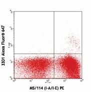
C57BL/6 splenocytes stained with M5/114 (I-A/I-E) PE and 33D1 Alexa Fluor® 647
| Cat # | Size | Price | Quantity Check Availability | Save | ||
|---|---|---|---|---|---|---|
| 124912 | 100 µg | 190€ | ||||
33D1 antigen, also known as DCIR2 (dendritic cell inhibitory receptor 2), is reported to be a dendritic cell marker on subpopulation DC in the spleen, thymus, and Peyer's patch. It is reported that most N418 (CD11c)+ cells in spleen are also 33D1 positive. Bone marrow dendritic cells upregulate 33D1 antigen expression after incubation with GM-CSF.
Product DetailsProduct Details
- Verified Reactivity
- Mouse
- Antibody Type
- Monoclonal
- Host Species
- Rat
- Immunogen
- Mouse dendritic cells
- Formulation
- Phosphate-buffered solution, pH 7.2, containing 0.09% sodium azide.
- Preparation
- The antibody was purified by affinity chromatography and conjugated with Alexa Fluor® 647 under optimal conditions.
- Concentration
- 0.5 mg/ml
- Storage & Handling
- The antibody solution should be stored undiluted between 2°C and 8°C, and protected from prolonged exposure to light. Do not freeze.
- Application
-
FC - Quality tested
- Recommended Usage
-
Each lot of this antibody is quality control tested by immunofluorescent staining with flow cytometric analysis. For flow cytometric staining, the suggested use of this reagent is ≤ 0.25 µg per 106 cells in 100 µl volume. It is recommended that the reagent be titrated for optimal performance for other applications.
* Alexa Fluor® 647 has a maximum emission of 668 nm when it is excited at 633nm / 635nm.
Alexa Fluor® and Pacific Blue™ are trademarks of Life Technologies Corporation.
View full statement regarding label licenses - Excitation Laser
-
Red Laser (633 nm)
- Application Notes
-
Additional reported applications (for the relevant formats) include: immunofluorescence3 and immunohistochemical staining of acetone-fixed frozen sections4.
Binding of clone 33D1 is calcium-dependent. We recommend staining in EDTA-free FACS buffer containing a source of Ca2+ ions.
- Application References
-
- Crowley MT, et al. 1990. J. Immunol. Methods 133:55.
- Nussenzwig MC, et al. 1982. P. Natl. Acad. Sci. USA 79:161.
- Steinman RM, et al. 1983. J. Exp. Med. 157:613. (IF)
- Fischer HG, et al. 2000. J. Immunol. 164:4826. (IHC)
- Product Citations
-
- RRID
-
AB_1227623 (BioLegend Cat. No. 124912)
Antigen Details
- Structure
- The antigen recognized by 33D1 is yet unknown.
- Distribution
-
33D1 antigen was reported on dendritic cell populations from mouse thymus, spleen lymph nodes and Peyer patches. Bone marrow dendritic cells can be induced to express 33D1 antigen in the presence of GM-CSF. In mouse spleen, most N418+ cells are also 33D1 positive.
- Function
- GM-CSF is reported to increase expression of 33D1 antigen on dendritic cells from bone marrow cells and IL-4 reported to down regulate the 33D1 antigen.
- Cell Type
- Dendritic cells
- Biology Area
- Immunology
- Molecular Family
- CD Molecules
- Gene ID
- NA
- UniProt
- View information about 33D1 on UniProt.org
Related Pages & Pathways
Pages
Related FAQs
Other Formats
View All 33D1 Reagents Request Custom Conjugation| Description | Clone | Applications |
|---|---|---|
| Purified anti-mouse DC Marker (33D1) | 33D1 | FC,IHC-F,ICC |
| Biotin anti-mouse DC Marker (33D1) | 33D1 | FC |
| PE anti-mouse DC Marker (33D1) | 33D1 | FC |
| FITC anti-mouse DC Marker (33D1) | 33D1 | FC |
| Alexa Fluor® 488 anti-mouse DC Marker (33D1) | 33D1 | FC |
| Alexa Fluor® 647 anti-mouse DC Marker (33D1) | 33D1 | FC |
| APC anti-mouse DC Marker (33D1) | 33D1 | FC |
Compare Data Across All Formats
This data display is provided for general comparisons between formats.
Your actual data may vary due to variations in samples, target cells, instruments and their settings, staining conditions, and other factors.
If you need assistance with selecting the best format contact our expert technical support team.
-
Purified anti-mouse DC Marker (33D1)
-
Biotin anti-mouse DC Marker (33D1)
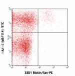
BALB/c splenocytes stained with I-A/I-E (M5/114) FITC and bi... -
PE anti-mouse DC Marker (33D1)
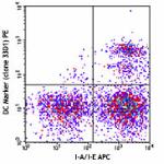
C57BL/6 splenocytes were stained with I-A/I-E APC and DC Mar... 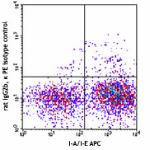
-
FITC anti-mouse DC Marker (33D1)
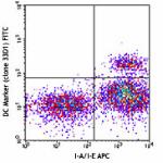
C57BL/6 splenocytes were stained with I-A/I-E APC and DC Mar... 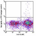
-
Alexa Fluor® 488 anti-mouse DC Marker (33D1)
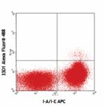
C57BL/6 splenocytes stained with I-A/I-E (M5/114.15.2) APC a... 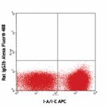
C57BL/6 splenocytes stained with I-A/I-E (M5/115) APC and Al... -
Alexa Fluor® 647 anti-mouse DC Marker (33D1)
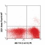
C57BL/6 splenocytes stained with M5/114 (I-A/I-E) PE and 33D... -
APC anti-mouse DC Marker (33D1)
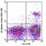
C57BL/6 splenocytes were stained with I-A/I-E PE and DC Mark... 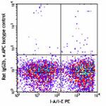
 Login / Register


Follow Us