 Login / Register


- Clone
- 177.19 (See other available formats)
- Regulatory Status
- RUO
- Other Names
- Autophagy protein 5
- Isotype
- Mouse IgG1, κ
- Ave. Rating
- Submit a Review
- Product Citations
- publications

-

Western blot of anti-ATG5 antibody (clone 177.19). The blot was incubated with the primary antibody at 0.2 µg/ml overnight at 4°C, followed by the incubation with horseradish peroxidase labeled goat anti-mouse secondary antibody (cat# 405306). Enhanced chemiluminescence was used as the detection system. Lysates: 10 µg. Recombinant human ATG5: 5 ng. -

ICC staining of anti-ATG5 antibody (clone 177.19) on Hela cells. The cells were fixed with 4% PFA, permeabilized with 0.1% Triton X-100, and blocked with 2% normal goat serum and 0.02%BSA. The cells were then stained with 0.5 µg/ml of the primary antibody overnight at 4°C, followed by incubation with Alexa Fluor® 594 goat anti-Mouse IgG (Cat. No. 405326) for one hour at room temperature. Cells were counterstained with Phalloidin™ Green 488 (Cat. No. 424201) and DAPI to visualize actin filaments and nuclei, respectively. The images were captured with a 60X objective. Scale bar: 50 µm. -

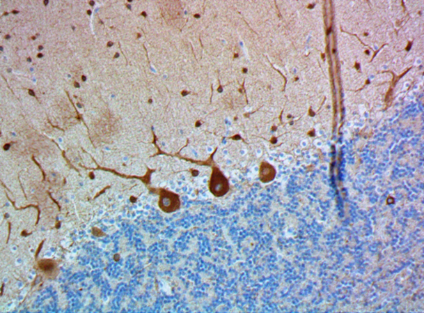
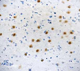
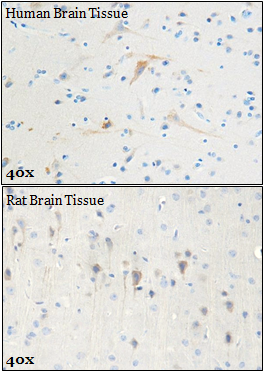
IHC staining of anti-ATG5 antibody (clone 177.19) on formalin-fixed paraffin-embedded normal human brain tissue. Following antigen retrieval using sodium citrate, the tissue was incubated with the primary antibody for 60 minutes at room temperature at 10 µg/ml. BioLegend's Ultra-Streptavidin (USA) HRP Detection Kit was used for detection. -

Western blot of purified anti-ATG5 antibody (clone 177.19). Lane 1: Molecular weight marker; Lane 2: 20 µg of Drosophila head lysate. The blot was incubated with 5 µg/mL of the primary antibody overnight at 4°C, followed by incubation with HRP-labeled goat anti-mouse IgG (Cat. No. 436302). Enhanced chemiluminescence was used as the detection system. -

ICC staining of purified anti-ATG5 antibody (clone 177.19) on Drosophila S2 cells. The cells were fixed with 4% PFA, permeabilized with a buffer containing 0.1% Triton X-100 and 0.25% BSA, and blocked with 2% normal goat serum and 0.02% BSA. The cells were then incubated with 1 µg/mL of the primary antibody overnight at 4°C, followed by incubation wit 2.5 µg/mL of Alexa Fluor® 647 goat anti-mouse IgG (Cat. No. 405322) for one hour at room temperature. Nuclei were counterstained with DAPI. The image was captured with a 60X objective. Scale bar: 20 µm
| Cat # | Size | Price | Quantity Check Availability | Save | ||
|---|---|---|---|---|---|---|
| 847401 | 25 µg | 90€ | ||||
| 847402 | 100 µg | 231€ | ||||
Autophagy protein 5 (ATG5) is involved in autophagic vesicle formation. ATG5 forms a complex with autophagy protein 12 (ATG12), which has E3 ubiquitin ligase activity, and is associated with autophagosome vesicle formation. ATG5 detaches from membranes immediately before or after autophagosome formation. ATG5 is also involved in mitochondrial quality control after oxidative damage, the innate anti-viral immune response, and apoptosis.
Product DetailsProduct Details
- Verified Reactivity
- Human, Mouse, Rat, Drosophila
- Antibody Type
- Monoclonal
- Host Species
- Mouse
- Immunogen
- ATG5 peptide conjugated to KLH.
- Formulation
- Phosphate-buffered solution, pH 7.2, containing 0.09% sodium azide.
- Preparation
- The antibody was purified by affinity chromatography.
- Concentration
- 0.5 mg/ml
- Storage & Handling
- The antibody solution should be stored undiluted between 2°C and 8°C.
- Application
-
WB - Quality tested
ICC, IHC-P - Verified
Direct ELISA - Reported in the literature, not verified in house - Recommended Usage
-
Each lot of this antibody is quality control tested by Western blotting. For Western blotting, the suggested use of this reagent is 0.1 - 1.0 µg/mL in human, mouse, and rat and 1.0 - 10 µg/mL in Drosophila For immunocytochemistry, a concentration range of 0.1 - 1.0 μg/mL is recommended in human, mouse, rat and 1.0 - 5.0 μg/mL in Drosophila. For immunohistochemical staining on formalin-fixed paraffin-embedded tissue sections, the suggested use of this reagent is 2.0 - 10 µg per mL. It is recommended that the reagent be titrated for optimal performance for each application.
- Application Notes
-
Reactivity to Drosophila was only verified with the purified format.
- RRID
-
AB_2734618 (BioLegend Cat. No. 847401)
AB_2734618 (BioLegend Cat. No. 847402)
Antigen Details
- Structure
- Human ATG5 is a 275 amino acid protein with a molecular mass of 32 kD.
- Distribution
-
Tissue distribution: Ubiquitous.
Cellular distribution: Vacuole, lysosome, and to a lesser extent mitochondria. - Function
- ATG5 is involved in many cellular processes including autophagic vesicle formation, mitochondrial quality control after oxidative damage, innate anti-viral immune response, and apoptosis.
- Interaction
- ATG12, LC3B, ATG16, TECPR1, Autophagy-linked FYVE protein.
- Biology Area
- Cell Biology, Neurodegeneration, Neuroscience, Neuroscience Cell Markers, Protein Trafficking and Clearance
- Molecular Family
- Autophagosome Markers
- Antigen References
-
1. Uchiyama Y, et al. 2008. Histochem. Cell Biol. 129:407. PubMed
2. Wu H, et al. 2014. Int. J. Biol. Sci. 10:1072. PubMed
3. Subramani S, Malhotra V. 2013. EMBO Rep. 14(2):143. PubMed - Gene ID
- 9474 View all products for this Gene ID
- UniProt
- View information about ATG5 on UniProt.org

Related FAQs
Other Formats
View All ATG5 Reagents Request Custom Conjugation| Description | Clone | Applications |
|---|---|---|
| Purified anti-ATG5 | 177.19 | WB,ICC,IHC-P,Direct ELISA |
| Alexa Fluor® 594 anti-ATG5 | 177.19 | ICC |
| Alexa Fluor® 647 anti-ATG5 | 177.19 | ICC |
| Spark YG™ 570 anti-ATG5 | 177.19 | ICC |
Customers Also Purchased
Compare Data Across All Formats
This data display is provided for general comparisons between formats.
Your actual data may vary due to variations in samples, target cells, instruments and their settings, staining conditions, and other factors.
If you need assistance with selecting the best format contact our expert technical support team.
-
Purified anti-ATG5

Western blot of anti-ATG5 antibody (clone 177.19). The blot ... 
ICC staining of anti-ATG5 antibody (clone 177.19) on Hela ce... 
IHC staining of anti-ATG5 antibody (clone 177.19) on formali... 
Western blot of purified anti-ATG5 antibody (clone 177.19). ... 
ICC staining of purified anti-ATG5 antibody (clone 177.19) o... -
Alexa Fluor® 594 anti-ATG5

ICC staining of Alexa Fluor® 594 anti-ATG5 antibody (clone 1... -
Alexa Fluor® 647 anti-ATG5

ICC staining of Alexa Fluor® 647 anti-ATG5 antibody (clone 1... -
Spark YG™ 570 anti-ATG5

HeLa cells were fixed with 1% paraformaldehyde (PFA) and blo...


Follow Us