- Clone
- DH8.3 (See other available formats)
- Regulatory Status
- RUO
- Other Names
- EGFRv3
- Isotype
- Mouse IgG1, κ
- Ave. Rating
- Submit a Review
- Product Citations
- publications

-

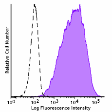
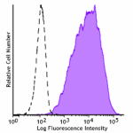
Human glioblastoma cell line DKMG/EGFRvIII was stained with purified anti-human EGFRvIII (clone DH8.3) (filled histogram) or purified mouse IgG1, κ isotype control (open histogram), followed by anti-mouse IgG PE. -

Whole cell extracts (15 µg total protein) from indicated cell lines resolved on a 4-12% Bis-Tris gel, transferred to a PVDF membrane, and probed with Purified anti-EGFRvIII antibody (clone DH8.3) overnight at 4°C. Proteins were visualized by chemiluminescence detection using HRP Goat anti-mouse IgG antibody (Cat. No. 405306) at a 1:3000 dilution. Direct-Blot™ HRP anti-β-actin antibody (Cat. No. 643807) was used as a loading control at a 1:10000 dilution. Lane M: Molecular weight marker
| Cat # | Size | Price | Quantity Check Availability | Save | ||
|---|---|---|---|---|---|---|
| 386002 | 100 µg | 349€ | ||||
Epidermal growth factor receptor variant III (EGFRvIII) is the result of an in frame deletion of exons 2-7 from extracellular region of EGFR, that results in the truncated receptor being constitutively active. This mutation is cancer-specific and is present in approximately 50% of glioblastoma tumors.
Product DetailsProduct Details
- Verified Reactivity
- Human
- Antibody Type
- Monoclonal
- Host Species
- Mouse
- Immunogen
- Peptide LEEKKGNYVVTDHC conjugated to KLH
- Formulation
- Phosphate-buffered solution, pH 7.2, containing 0.09% sodium azide
- Preparation
- The antibody was purified by affinity chromatography.
- Concentration
- 0.5 mg/mL
- Storage & Handling
- The antibody solution should be stored undiluted between 2°C and 8°C.
- Application
-
FC - Quality tested
WB - Verified
IHC-P - Reported in the literature, not verified in house - Recommended Usage
-
Each lot of this antibody is quality control tested by immunofluorescent staining with flow cytometric analysis. For flow cytometric staining, the suggested use of this reagent is ≤ 1.0 µg per million cells in 100 µL volume. For western blotting, the suggested use of this reagent is 0.125 - 1.0 µg/mL. It is recommended that the reagent be titrated for optimal performance for each application.
- Application References
-
- Banisadr A, et al. 2020. J Cell Sci. 133:jcs247189 (FC)
- Achim A, et al. 2003. Proc Natl Acad Sci U S A. 100:2163 (IHC, WB)
- Öhman L, et al. 2002. Tumor Biol. 23:61 (IHC)
- RRID
-
AB_3097613 (BioLegend Cat. No. 386002)
Antigen Details
- Structure
- Deletion of exons 2-7 of the EGFR gene. Molecular mass of approximately 145 kD
- Distribution
-
Present in approximately 50% of glioblastoma tumors
- Function
- Pro-tumorigenic (promotes tumor growth, survival, invasion, stemness, metabolism, and angiogenesis)
- Biology Area
- Cancer Biomarkers
- Antigen References
-
- Hui G, et al. 2013. FEBS Journal. 280:21.
- Gene ID
- 1956 View all products for this Gene ID
- UniProt
- View information about EGFRvIII on UniProt.org
Related FAQs
Other Formats
View All EGFRvIII Reagents Request Custom Conjugation| Description | Clone | Applications |
|---|---|---|
| Purified anti-human EGFRvIII | DH8.3 | FC,WB |
Compare Data Across All Formats
This data display is provided for general comparisons between formats.
Your actual data may vary due to variations in samples, target cells, instruments and their settings, staining conditions, and other factors.
If you need assistance with selecting the best format contact our expert technical support team.

 Login / Register


Follow Us