- Clone
- NSE-P2 (See other available formats)
- Regulatory Status
- RUO
- Other Names
- Gamma-enolase, neural enolase, neuron-specific enolase, 2-phospho-D-glycerate hydrolyase, ENO2, NSE
- Previously
-
Covance Catalog# MMS-519P
- Isotype
- Mouse IgG1, κ
- Ave. Rating
- Submit a Review
- Product Citations
- publications
-

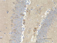
IHC staining of purified anti-NSE antibody (clone NSE-P2) on formalin-fixed paraffin-embedded mouse brain tissue. Following antigen retrieval using Sodium Citrate H.I.E.R., the tissue was incubated with 1 µg/ml of the primary antibody for 60 minutes at room temperature. BioLegend's Ultra-Streptavidin (USA) HRP kit (Multi-Species, DAB, Cat. No. 929901) was used for detection followed by hematoxylin counterstaining, according to the protocol provided. The image was captured with a 40X objective. Scale bar: 50 µm -

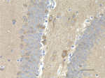
IHC staining of purified anti-NSE antibody (clone NSE-P2) on formalin-fixed paraffin-embedded rat brain tissue. Following antigen retrieval using Sodium Citrate H.I.E.R., the tissue was incubated with 1 µg/ml of the primary antibody for 60 minutes at room temperature. BioLegend's Ultra-Streptavidin (USA) HRP kit (Multi-Species, DAB, Cat. No. 929901) was used for detection followed by hematoxylin counterstaining, according to the protocol provided. The image was captured with a 40X objective. Scale bar: 50 µm -

IHC staining of purified anti-NSE antibody (clone NSE-P2) on formalin-fixed paraffin-embedded human pancreas tissue. Following antigen retrieval using Sodium Citrate H.I.E.R., the tissue was incubated with 10 µg/ml of the primary antibody overnight at 4°C . BioLegend's Ultra-Streptavidin (USA) HRP kit (Multi-Species, DAB, Cat. No. 929901) was used for detection followed by hematoxylin counterstaining, according to the protocol provided. The image was captured with a 10X objective. Scale bar: 50 µm -

Western blot of purified anti-NSE antibody (clone NSE-P2). Lane 1: Molecular weight marker; Lane 2: 20 µg of human brain lysate; Lane 3: 20 µg of mouse brain lysate; Lane 4: 20 µg of rat brain lysate. The blot was incubated with 5 µg/mL of the primary antibody overnight at 4°C, followed by incubation with HRP labeled goat anti-mouse IgG (Cat. No. 405306). Enhanced chemiluminescence was used as the detection system. -

ICC staining of purified anti-NSE antibody (clone NSE-P2) on SH-SY5Y Cells. The cells were fixed with 4% PFA, permeabilized with a buffer containing 0.1% Triton X-100 and 0.25% BSA, and blocked with 2% normal goat serum and 0.02% BSA. The cells were then incubated with 0.5 µg/mL of the primary antibody overnight at 4°C, followed by incubation with 2.5 µg/mL of Alexa Fluor® 594 goat anti-mouse IgG for one hour at room temperature. The cells were co-stained with Flash Phalloidin™ Green 488 (Cat. No. 424201). The slide was mounted with fluoromount G with DAPI. The image was captured with a 60X objective. Scale bar: 20 µm
| Cat # | Size | Price | Quantity Check Availability | Save | ||
|---|---|---|---|---|---|---|
| 804501 | 200 µL | 285€ | ||||
Neuron specific enolase is found in elevated concentrations in plasma and certain neoplasias. These include pediatric neuroblastoma and small cell lung cancer.
Neuron specific enolase (NSE) converts 2-phosphoglycerate to phosphoenolpyruvate in the glycolytic pathway, and the reverse reaction in gluconeogenesis. It is one of three mammalian enolases, which are also known as ENO1, ENO2, and ENO3 or as enolase alpha, beta and gamma. The three enolases have different cell type specific expression patterns, so that antibodies to them are useful cell type specific markers. NSE corresponds to ENO2 or enolase gamma and is heavily expressed in neuronal cells. Neurons require a great deal of energy and have high levels of glycolytic enzymes such as NSE. Antibodies to this NSE are useful for identifications of neuronal cell bodies, developing neuronal lineage and neuroendocrine cells. Release of NSE from damaged neurons into CSF and blood has also been used as a biomarker of neuronal injury.
Product Details
- Verified Reactivity
- Human, Mouse, Rat
- Antibody Type
- Monoclonal
- Host Species
- Mouse
- Immunogen
- This antibody was raised against a synthetic peptide corresponding to amino acids 271-285. It recognizes the sequence TGDQLGALYQDFVRD.
- Formulation
- Phosphate-buffered solution + 0.03% thimerosal.
- Preparation
- The antibody was purified by affinity chromatography.
- Concentration
- 1 mg/mL
- Storage & Handling
- The antibody solution should be stored undiluted between 2°C and 8°C. Please note the storage condition for this antibody has been changed from -20°C to between 2°C and 8°C. You can also check your vial or your CoA to find the most accurate storage condition for this antibody.
- Application
-
IHC-P - Quality tested
WB, ICC - Verified - Recommended Usage
-
Each lot of this antibody is quality control tested by formalin-fixed paraffin-embedded immunohistochemical staining. For immunohistochemistry, a concentration range of 1.0 - 10 µg/mL is suggested. For Western blotting, the suggested use of this reagent is 1.0 - 5.0 µg per mL. For immunocytochemistry, a concentration range of 0.5 - 10 μg/mL is recommended. It is recommended that the reagent be titrated for optimal performance for each application.
- RRID
-
AB_2564666 (BioLegend Cat. No. 804501)
Antigen Details
- Structure
- Expected MW: 47 kD
- Cell Type
- Mature Neurons, Neural Stem Cells
- Biology Area
- Cell Biology, Neuroscience, Neuroscience Cell Markers, Stem Cells
- Molecular Family
- Phospho-Proteins
- Antigen References
-
- Murray GI, et al. 1993. J Clin Pathol. 46:993.
- Duncan ME, et al. 1992. J immunol Methods. 151:227
- Gene ID
- 2026 View all products for this Gene ID
- UniProt
- View information about NSE on UniProt.org
Related Pages & Pathways
Pages
Other Formats
View All NSE Reagents Request Custom Conjugation| Description | Clone | Applications |
|---|---|---|
| Purified anti-NSE | NSE-P2 | IHC-P,WB,ICC |
Compare Data Across All Formats
This data display is provided for general comparisons between formats.
Your actual data may vary due to variations in samples, target cells, instruments and their settings, staining conditions, and other factors.
If you need assistance with selecting the best format contact our expert technical support team.
-
Purified anti-NSE
IHC staining of purified anti-NSE antibody (clone NSE-P2) on... 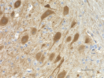
IHC staining of purified anti-NSE antibody (clone NSE-P2) on... 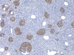
IHC staining of purified anti-NSE antibody (clone NSE-P2) on... 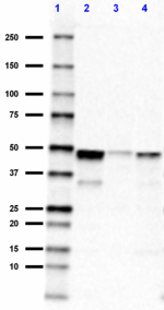
Western blot of purified anti-NSE antibody (clone NSE-P2). ... 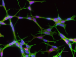
ICC staining of purified anti-NSE antibody (clone NSE-P2) on...

 Login / Register


Follow Us