 Login / Register


- Clone
- 10D1 (See other available formats)
- Regulatory Status
- RUO
- Other Names
- Structure specific recognition protein 1 (SSRP1), Facilitates Chromatin Transcription 80 kD subunit, FACTp80, Recombination signal sequence recognition protein
- Isotype
- Mouse IgG2b, κ
- Ave. Rating
- Submit a Review
- Product Citations
- publications

-

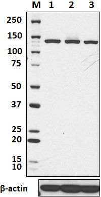
Total cell lysates (15 µg total protein) from THP-1, HaCat, Alexa Fluor® 594 and NTERA2 cells were resolved by 4-12% Bis-Tris gel electrophoresis, transferred to a nitrocellulose membrane, and probed with 1.0 µg/mL of purified anti-SSRP1 antibody, clone 10D1, overnight at 4°C. Proteins were visualized by chemiluminescence detection using HRP goat anti-mouse IgG antibody (Cat. No. 405306) at a 1:3000 dilution. Direct-Blot™ HRP anti-β-actin antibody (Cat. No. 664804) was used as a loading control at a 1:10000 dilution (lower). Lane M: Molecular Weight marker. -

Total lysates (15 µg protein) from HeLa (lane 1) and NIH3T3 cells (lane 2) were resolved by electrophoresis (4-12% Bis-Tris gel), transferred to nitrocellulose, and probed with 1:500 diluted (1 µg/mLPurified anti-SSRP1 Antibody, clone 10D1 (upper) or 1:3000 diluted Purified anti-β-actin Antibody, clone Poly6221 (lower). Proteins were visualized by chemiluminescence detection using a 1:3000 diluted goat anti-mouse-IgG secondary antibody conjugated to HRP for the anti-SSRP1 Antibody, and a donkey anti-rabbit IgG Antibody conjugated to HRP for anti-β-actin Antibody. -

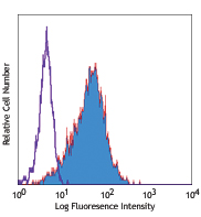
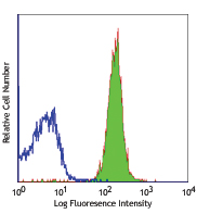
HeLa cells were stained with purified anti- SSRP-1 (10D1) antibody, followed by staining with DyLight™ 594 conjugated goat anti-mouse IgG (red) antibody. Actin filaments were labeled in green. Nuclei were stained with DAPI (blue). -

Chromatin Immunoprecipitations (ChIP) were performed with cross-linked chromatin samples from 4 X 10⁶ HeLa cells (starved overnight then treated with 10% FCS) with either A) 1:50 dilution of Go-ChIP-Grade™ Purified anti-SSRP1 Antibody (clone 10D1, Cat. No. 609709) or B) equal amount of Purified Mouse IgG2b, κ isotype control antibody (clone MPC-11, Cat. No. 400301) by using Go-ChIP-Grade™ Protein G Enzymatic Kit (Cat. No. 699904). The enriched DNA was purified and quantified by real-time qPCR using primers targeting human EGR1 gene region or α-Satellite repeats. The amount of immunoprecipitated DNA in each sample is represented as signal relative to total amount of input chromatin.
| Cat # | Size | Price | Quantity Check Availability | Save | ||
|---|---|---|---|---|---|---|
| 609701 | 25 µg | 81€ | ||||
| 609702 | 100 µg | 175€ | ||||
SSRP1 is a nuclear protein and a component of the FACT complex (facilitates chromatin transcription). The FACT complex is composed of the SUPT16H (FACT140) and the SSRP1 FACTp80 proteins. This complex interacts with nucleosomes and histone H2A/H2B dimers to promote nucleosome disassembly and allow transcription elongation. This protein interacts with dsDNA. The SSRP1 protein has been shown to be modified by cisplatin and may protect DNA from repair and block DNA replication. The 10D1 monoclonal antibody recognizes the human SSRP1 protein and has been shown to be useful for Western blotting.
Product DetailsProduct Details
- Verified Reactivity
- Human, Mouse
- Antibody Type
- Monoclonal
- Host Species
- Mouse
- Immunogen
- Recombinant (partial) (308-524aa)
- Formulation
- This antibody is provided in phosphate-buffered solution, pH 7.2, containing 0.09% sodium azide at 0.5 mg/ml.
- Preparation
- The antibody was purified by affinity chromatography.
- Concentration
- 0.5 mg/ml
- Storage & Handling
- Upon receipt, store undiluted between 2°C and 8°C.
- Application
-
WB - Quality tested
ICC, ChIP - Verified
IP - Reported in the literature, not verified in house - Recommended Usage
-
Each lot of this antibody is quality control tested by Western blotting. Western blotting, suggested working dilution(s): Use 5 µg per 5 ml antibody dilution buffer for each mini-gel. For ChIP applications, the suggested dilution is 1:50-1:200 by volume. It is recommended that the reagent be titrated for optimal performance for each application.
- Application Notes
-
10D1 displayed a stronger affinity for SSRP1 than BioLegend clone 3E4 when tested for western blot application.
- Application References
-
- Tan BC, et al. 2006. EMBO 25:3975. (ChIP)
- Carvalho S, et al. 2013. Nucleic Acids Res. 41:2881. PubMed
- Tan BC, et al. 2004. J. Biol. Chem. 279:9321. (Immunogen)
- Lolis AA, et al. 2013. J Biol Chem. 288:7676. PubMed
- Aida M, et al. 2013. PNAS. 110:7784. PubMed
- Dermawan JK, et al. 2014. Mol Cancer Ther. 13:2203. PubMed
- Shakya A, et al. 2015. Mol Cell Biol. 35:1014. PubMed
- Product Citations
-
- RRID
-
AB_315730 (BioLegend Cat. No. 609701)
AB_315731 (BioLegend Cat. No. 609702)
Antigen Details
- Structure
- SSRP family, HMG box domain; 81 kD
- Distribution
-
Nuclear
- Function
- Interacts with nucleosomes and histone H2A/H2B dimers to promote nucleosome disassembly and allow transcription elongation, binds to dsDNA. Modified by cisplatin, may protect DNA from repair and block DNA replication
- Interaction
- Component of FACT ATP-dependent chromatin remodeling complex, SUPT16H/Cdc68 in FACT complex, H2A/H2B dimers
- Biology Area
- Cell Biology, Chromatin Remodeling/Epigenetics, DNA Repair/Replication, Transcription Factors
- Antigen References
-
1. LeRoy G, et al. 1998. Science 282:1836.
2. Orphanides G, et al. 1998. Cell 92:105.
3. Orphanides G, et al. 1999. Nature 400:284. - Gene ID
- 6749 View all products for this Gene ID
- UniProt
- View information about SSRP1 on UniProt.org

Related FAQs
Other Formats
View All SSRP1 Reagents Request Custom Conjugation| Description | Clone | Applications |
|---|---|---|
| Purified anti-SSRP1 | 10D1 | WB,ICC,IP,ChIP |
Customers Also Purchased
Compare Data Across All Formats
This data display is provided for general comparisons between formats.
Your actual data may vary due to variations in samples, target cells, instruments and their settings, staining conditions, and other factors.
If you need assistance with selecting the best format contact our expert technical support team.


Follow Us