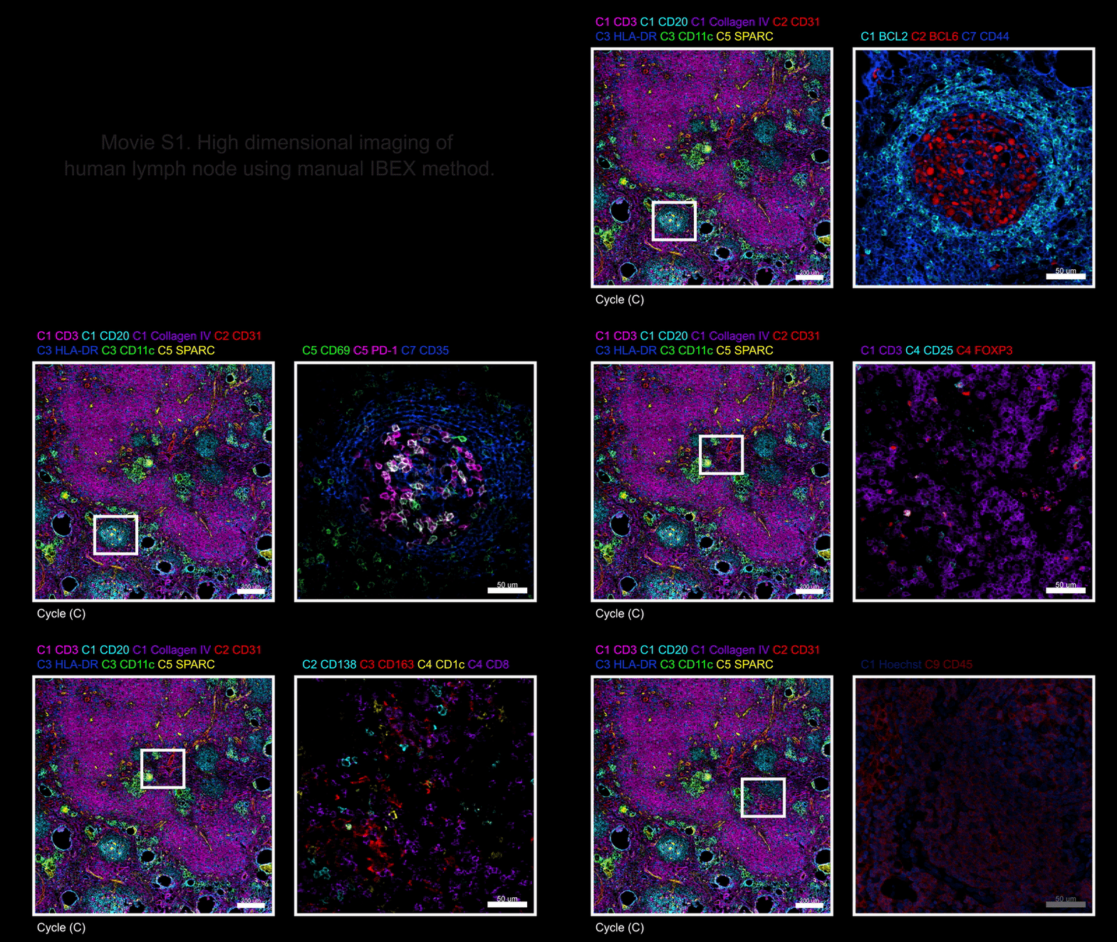
Highly-multiplexed imaging technologies like IBEX enable the simultaneous detection of dozens of cellular features on a single piece of tissue. Below is a representative 38-plex dataset generated with the IBEX technique originally published in Nature Protocols, using tissue sections from a single human lymph node. The images reveal the high level of detail achieved at both low and high magnification for groupings of related cellular and tissue markers. This dataset, including details on the 25 BioLegend reagents used in its creation, along with others are also available open-access through the authors’ Zenodo affiliate publication.
For additional questions on getting started with IBEX, reference our IBEX FAQs.
IBEX Data
Watch the images taken in the Nature Protocols 38-plex data set cycle through or watch the full video.
Webinars
This 2022 on-demand webinar by the developers of the IBEX technology discusses how antibody-based imaging empowers the study of dozens of protein biomarkers in a variety of tissue sections. Learn how they analyze normal and malignant lymph nodes, optimize tissue processing of fixed frozen and FFPE samples, test antibody validation, and create organ mapping antibody panels.
This initial 2021 webinar focuses on the development of IBEX (Iterative Bleaching Extended multi-pleXity) imaging method—an open microscopy method for high content multiplex imaging of diverse tissues. Andrea J. Radtke, Ph.D. of the Ronald Germain Lab at the Center for Advanced Tissue Imaging, NIAID/NIH discusses the method and its applications in cell atlas building and tissue phenotyping. IBEX allows for repeated cycles of staining, imaging, and bleaching, conveniently using hundreds of commercially available antibodies and enabling the analysis of more than 65 parameters in a single sample.
 Login / Register


Follow Us