- Clone
- Poly28410 (See other available formats)
- Regulatory Status
- RUO
- Other Names
- 160 kD Neurofilament Protein, Neurofilament Triplet M Protein, Neurofilament-3 (150 Kd Medium), Neurofilament, Medium Polypeptide 150kD
- Previously
-
Covance Catalog# PRB-575C
- Isotype
- Rabbit Polyclonal IgG
- Ave. Rating
- Submit a Review
- Product Citations
- publications
-

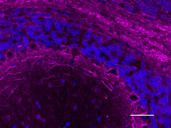
IHC staining of anti-Neurofilament M (NF-M) antibody (Poly28410) on formalin-fixed paraffin-embedded mouse cerebellum tissue. Following antigen retrieval using Sodium Citrate H.I.E.R., the tissue was incubated with a 1:200 dilution of the primary antibody overnight at 4°C, followed by incubation with Alexa Fluor® 647 donkey anti-rabbit IgG (Cat. No. 406414) for one hour at room temperature. The slide was mounted with Fluoromount-G with DAPI. The image was captured with a 40X objective. Scale Bar: 50 µm -

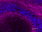
IHC staining of anti-Neurofilament M (NF-M) antibody (Poly28410) on formalin-fixed paraffin-embedded rat cerebellum tissue. Following antigen retrieval using Sodium Citrate H.I.E.R., the tissue was incubated with a 1:200 dilution of the primary antibody overnight at 4°C, followed by incubation with Alexa Fluor® 647 donkey anti-rabbit IgG (Cat. No. 406414) for one hour at room temperature. The slide was mounted with Fluoromount-G with DAPI. The image was captured with a 40X objective. Scale Bar: 50 µm -

IHC staining of anti-Neurofilament M (NF-M) antibody (Poly28410) on formalin-fixed paraffin-embedded human cortex tissue. Following antigen retrieval using Sodium Citrate H.I.E.R., the tissue was incubated with a 1:200 dilution of the primary antibody overnight at 4°C, followed by incubation with Alexa Fluor® 647 donkey anti-rabbit IgG (Cat. No. 406414) for one hour at room temperature. The slide was mounted with Fluoromount-G with DAPI. The image was captured with a 40X objective. Scale Bar: 50 µm -

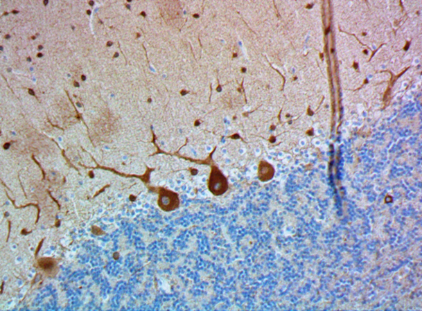
IHC staining of anti-Neurofilament M (NF-M) antibody (Poly28410) on formalin-fixed paraffin-embedded rat brain tissue. Following antigen retrieval using Sodium Citrate H.I.E.R., the tissue was incubated with a 1:500 dilution of the primary antibody for 20 minutes at room temperature. BioLegend's Ultra-Streptavidin (USA) HRP kit (Multi-Species, DAB, Cat. No. 929901) was used for detection followed by hematoxylin counterstaining, according to the protocol provided. The image was captured with a 40X objective. Scale bar: 50 µm -

Western blot of anti-Neurofilament M (NF-M) antibody (Poly28410). Lane 1: Molecular weight marker; Lane 2: 30 µg of human brain lysate; Lane 3: 30 µg of mouse brain lysate; Lane 4: 30 µg of rat brain lysate. The blot was incubated with a 1:10,000 dilution of the primary antibody overnight at 4°C, followed by incubation with HRP anti-rabbit IgG (Cat. No. 410406). Enhanced chemiluminescence was used as the detection system.
| Cat # | Size | Price | Quantity Check Availability | Save | ||
|---|---|---|---|---|---|---|
| 841001 | 100 µL | 251€ | ||||
Neurofilaments can be defined as the intermediate or 10nm diameter filaments found in neuronal cells. They are composed a mixture of subunits which often includes the neurofilament triplet proteins, NF-L, NF-M and NF-H. Neurofilaments may also include peripherin, a-internexin, nestin and in some cases vimentin. Antibodies in the various neurofilament subunits are very useful cell type markers since the proteins are quite abundant, biochemically stable.
Product DetailsProduct Details
- Verified Reactivity
- Human, Rat, Mouse
- Antibody Type
- Polyclonal
- Host Species
- Rabbit
- Immunogen
- The immunogen was the extreme carboxyterminal region of rat NF-M, which was expressed in E. coli and purified by ion exchange chromatograpy.
- Preparation
- Serum
- Storage & Handling
- Store at -20°C. Upon initial thawing, apportion into working aliquots and store at -20°C. Avoid repeated freeze-thaw cycles to prevent denaturing the antibody.
- Application
-
IHC-P - Quality tested
WB - Verified - Recommended Usage
-
Each lot of this antibody is quality control tested by formalin-fixed paraffin-embedded immunohistochemical staining. For immunohistochemistry, a dilution range of 1:200 - 1:500 is suggested. For Western blotting, the suggested use of this reagent is 1:5000 - 1:10000. It is recommended that the reagent be titrated for optimal performance for each application.
- Application Notes
-
The production and characterization of this antiserum similar to monoclonal NF-M clone 3H11.
This product may contain other non-IgG subtypes. - Application References
-
- Harris J, Ayyub C, Shaw G. A molecular dissection of the carboxyterminal tails of the major neurofilament subunits NF-M and NF-H. J. Neurosci. Res. 30:47-62, 1991.
- Product Citations
-
- RRID
-
AB_2565457 (BioLegend Cat. No. 841001)
Antigen Details
- Structure
- Expected MW: 145-160 kD
- Cell Type
- Mature Neurons
- Biology Area
- Cell Biology, Neuroscience, Neuroscience Cell Markers
- Molecular Family
- Intermediate Filaments
- Gene ID
- 4741 View all products for this Gene ID
- UniProt
- View information about Neurofilament M on UniProt.org
Related Pages & Pathways
Pages
Related FAQs
Other Formats
View All Neurofilament M (NF-M) Reagents Request Custom Conjugation| Description | Clone | Applications |
|---|---|---|
| Anti-Neurofilament M (NF-M) | Poly28410 | IHC-P,WB |
Customers Also Purchased
Compare Data Across All Formats
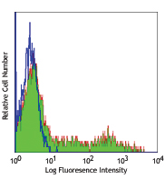
This data display is provided for general comparisons between formats.
Your actual data may vary due to variations in samples, target cells, instruments and their settings, staining conditions, and other factors.
If you need assistance with selecting the best format contact our expert technical support team.
-
Anti-Neurofilament M (NF-M)
IHC staining of anti-Neurofilament M (NF-M) antibody (Poly28... 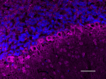
IHC staining of anti-Neurofilament M (NF-M) antibody (Poly28... 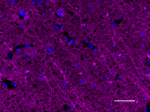
IHC staining of anti-Neurofilament M (NF-M) antibody (Poly28... 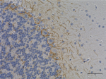
IHC staining of anti-Neurofilament M (NF-M) antibody (Poly28... 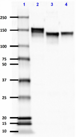
Western blot of anti-Neurofilament M (NF-M) antibody (Poly28...
 Login / Register




Follow Us