- Clone
- A17188B (See other available formats)
- Regulatory Status
- RUO
- Other Names
- PD1, PDCD1
- Isotype
- Mouse IgG2b, κ
- Ave. Rating
- Submit a Review
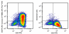
| Cat # | Size | Price | Quantity Check Availability | Save | ||
|---|---|---|---|---|---|---|
| 621619 | 25 tests | 151€ | ||||
| 621620 | 100 tests | 315€ | ||||
Programmed cell death protein 1 (PD-1), also known as CD279, is a 55 kD member of the immunoglobulin superfamily. CD279 contains the immunoreceptor tyrosine-based inhibitory motif (ITIM) in the cytoplasmic region and plays a key role in peripheral tolerance and autoimmune disease. CD279 is expressed predominantly on activated T cells, B cells, and myeloid cells. PD-L1 (B7-H1, CD274) and PD-L2 (B7-DC, CD273) are ligands of CD279 (PD-1) and are members of the B7 gene family. Evidence suggests overlapping functions for these two PD-1 ligands and their constitutive expression on some normal tissues and upregulation on activated antigen-presenting cells. Interaction of CD279 ligands results in inhibition of T cell proliferation and cytokine secretion.
Product DetailsProduct Details
- Verified Reactivity
- Human
- Antibody Type
- Monoclonal
- Host Species
- Mouse
- Immunogen
- Recombinant human CD279 protein
- Formulation
- Phosphate-buffered solution, pH 7.2, containing 0.09% sodium azide and BSA (origin USA)
- Preparation
- The antibody was purified by affinity chromatography and conjugated with APC/Fire™ 810 under optimal conditions.
- Concentration
- Lot-specific (to obtain lot-specific concentration and expiration, please enter the lot number in our Certificate of Analysis online tool.)
- Storage & Handling
- The antibody solution should be stored undiluted between 2°C and 8°C, and protected from prolonged exposure to light. Do not freeze.
- Application
-
FC - Quality tested
- Recommended Usage
-
Each lot of this antibody is quality control tested by immunofluorescent staining with flow cytometric analysis. For flow cytometric staining, the suggested use of this reagent is 5 µL per million cells in 100 µL staining volume or 5 µL per 100 µL of whole blood. It is recommended that the reagent be titrated for optimal performance for each application.
* APC/Fire™ 810 has a maximum excitation of 650 nm and a maximum emission of 810 nm.
Excessive exposure to light, and commonly used fixation, permeabilization buffers can affect APC/Fire™ 810 fluorescence signal intensity and spread. Please keep conjugates protected from light exposure. For more information and representative data, visit our Fire Dyes page. - Excitation Laser
-
Red Laser (633 nm)
- Application Notes
-
A17188B antibody can block the binding of NAT105 and EH12.2H7 antibodies to the target.
- RRID
-
AB_2910488 (BioLegend Cat. No. 621619)
AB_2910488 (BioLegend Cat. No. 621620)
Antigen Details
- Structure
- 55 kD type I transmembrane protein
- Distribution
-
Transiently expressed on CD4- and CD8- thymocytes, upregulated in thymocytes and splenic T and B lymphocytes, and is expressed on activated myeloid cells.
- Function
- Signaling and co-stimulation (co-inhibition)
- Interaction
- SHP-1 and SHP-2
- Ligand/Receptor
- PD-L1 (CD274) and PD-L2 (CD273)
- Cell Type
- Lymphocytes, T cells
- Biology Area
- Cell Biology, Immuno-Oncology, Inhibitory Molecules
- Molecular Family
- CD Molecules
- Antigen References
-
- Ishida Y, et al. 1992. EMBO J. 11:3887
- Francisco LM, et al. 2010. Immunol Rev. 236:219
- Gene ID
- 5133 View all products for this Gene ID
- UniProt
- View information about CD279 on UniProt.org
Related FAQs
Other Formats
View All CD279 Reagents Request Custom Conjugation| Description | Clone | Applications |
|---|---|---|
| Ultra-LEAF™ Purified anti-human CD279 (PD-1) | A17188B | Block |
| Purified anti-human CD279 (PD-1) | A17188B | FC,Direct ELISA |
| PE anti-human CD279 (PD-1) | A17188B | FC |
| APC anti-human CD279 (PD-1) | A17188B | FC |
| FITC anti-human CD279 (PD-1) | A17188B | FC |
| PerCP/Cyanine5.5 anti-human CD279 (PD-1) | A17188B | FC |
| PE/Cyanine7 anti-human CD279 (PD-1) | A17188B | FC |
| TotalSeq™-D1169 anti-human CD279 (PD-1) | A17188B | PG |
| APC/Fire™ 810 anti-human CD279 (PD-1) | A17188B | FC |
| PE/Fire™ 640 anti-human CD279 (PD-1) | A17188B | FC |
| PE/Fire™ 700 anti-human CD279 (PD-1) | A17188B | FC |
| PE/Fire™ 810 anti-human CD279 (PD-1) | A17188B | FC |
| Biotin anti-human CD279 (PD-1) | A17188B | Direct ELISA |
| PE/Dazzle™ 594 anti-human CD279 (PD-1) | A17188B | FC |
| KIRAVIA Blue 520™ anti-human CD279 (PD-1) | A17188B | FC |
| Spark Red™ 718 anti-human CD279 (PD-1) | A17188B | FC |
Customers Also Purchased
Compare Data Across All Formats
This data display is provided for general comparisons between formats.
Your actual data may vary due to variations in samples, target cells, instruments and their settings, staining conditions, and other factors.
If you need assistance with selecting the best format contact our expert technical support team.
-
Ultra-LEAF™ Purified anti-human CD279 (PD-1)
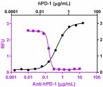
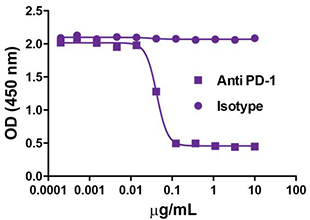
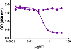
Recombinant hPD-1 binds to hPD-L1. Anti-human PD-1 Antibody ... -
Purified anti-human CD279 (PD-1)

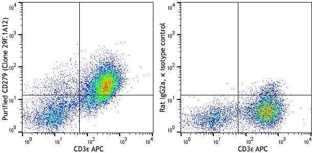
PHA-stimulated (3 days) human peripheral blood lymphocytes w... 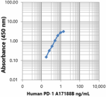
Direct ELISA showing purified anti-human PD-1 (clone A17188B... -
PE anti-human CD279 (PD-1)

PHA-stimulated (day-3) human peripheral blood lymphocytes we... -
APC anti-human CD279 (PD-1)

PHA-stimulated (day-3) human peripheral blood lymphocytes we... -
FITC anti-human CD279 (PD-1)

PHA-stimulated (day-3) human peripheral blood lymphocytes we... -
PerCP/Cyanine5.5 anti-human CD279 (PD-1)

PHA-stimulated (day-3) human peripheral blood lymphocytes we... -
PE/Cyanine7 anti-human CD279 (PD-1)

PHA-stimulated (day-3) human peripheral blood lymphocytes we... -
TotalSeq™-D1169 anti-human CD279 (PD-1)
-
APC/Fire™ 810 anti-human CD279 (PD-1)

PHA-stimulated (three days) human peripheral blood lymphocyt... -
PE/Fire™ 640 anti-human CD279 (PD-1)

PHA-stimulated (three days) human peripheral blood lymphocyt... -
PE/Fire™ 700 anti-human CD279 (PD-1)

PHA-stimulated (three days) human peripheral blood lymphocyt... -
PE/Fire™ 810 anti-human CD279 (PD-1)

PHA-stimulated (three days) human peripheral blood lymphocyt... -
Biotin anti-human CD279 (PD-1)
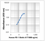
Direct ELISA showing biotin anti-human PD-1 (clone A17188B) ... -
PE/Dazzle™ 594 anti-human CD279 (PD-1)

PHA-stimulated (three days) human peripheral blood lymphocyt... -
KIRAVIA Blue 520™ anti-human CD279 (PD-1)

PHA-stimulated (three days) human peripheral blood lymphocyt... -
Spark Red™ 718 anti-human CD279 (PD-1)

Human peripheral blood lymphocytes were stained with anti-hu...

 Login / Register





Follow Us