- Clone
- Charlotte d1.23 (See other available formats)
- Regulatory Status
- RUO
- Other Names
- Retinoid acid early inducible gene-1δ, RAE-1δ, RAE-1δ
- Isotype
- Mouse IgG1
- Ave. Rating
- Submit a Review
- Product Citations
- publications
-

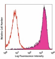
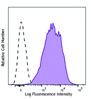
RAE-1δ transfected cells stained with Charlotte d1.23 PE
| Cat # | Size | Price | Quantity Check Availability | Save | ||
|---|---|---|---|---|---|---|
| 133203 | 50 µg | 98€ | ||||
RAE-1δ is one of the five RAE-1 family, GPI-linked membrane protein consisting of alpha, beta, gamma, delta, and epsilon. They are strong homology within the family, related by 92%-95% sequence identity. They are distantly related to MHC class I proteins. RAE-1 proteins are abundantly expressed in fetal tissues, but not in normal adult tissue. They are constitutively expressed on some tumors and can be induced by retinoid acid. Its ligand NKG2D is found on NK cells, many transformed cell lines, and LPS-stimulated peritoneal macrophages. The interaction of NKG2D with RAE-1 transmits stimulatory signals to NK cells and other hematopoietic cells, leading to enhanced proliferation, cytokine secretion and target killing. They are involved in the regulation of innate and immune cytotoxic responses to tumors, pathogen-infected cells, and autoimmune diseases.
Product DetailsProduct Details
- Verified Reactivity
- Mouse
- Antibody Type
- Monoclonal
- Host Species
- Mouse
- Immunogen
- Mouse RAE-1δ transfected RMA-S cell line
- Formulation
- Phosphate-buffered solution, pH 7.2, containing 0.09% sodium azide.
- Preparation
- The antibody was purified by affinity chromatography, and conjugated with PE under optimal conditions.
- Concentration
- 0.2 mg/ml
- Storage & Handling
- The antibody solution should be stored undiluted between 2°C and 8°C, and protected from prolonged exposure to light. Do not freeze.
- Application
-
FC - Quality tested
- Recommended Usage
-
Each lot of this antibody is quality control tested by immunofluorescent staining with flow cytometric analysis. For flow cytometric staining, the suggested use of this reagent is ≤0.015 µg per million cells in 100 µl volume. It is recommended that the reagent be titrated for optimal performance for each application.
- Excitation Laser
-
Blue Laser (488 nm)
Green Laser (532 nm)/Yellow-Green Laser (561 nm)
- Application References
-
- Lodoen M, et al. 2003. J. Exp. Med. 197:1245
- Carayannopoulos LN, et al. 2002. Eur J. Immunol.32 (3):597
- RRID
-
AB_1595625 (BioLegend Cat. No. 133203)
Antigen Details
- Structure
- RAE is a family of GPI-linked membrane protein consisting of alpha, beta, gamma, delta, and epsilon. They are strong homology within the family, related by 92%-95% sequence identity. They are distantly related to MHC class I proteins.
- Distribution
- RAE-1 proteins are abundantly expressed in fetal tissues, but not in normal adult tissue. They are constitutively expressed on some tumors and can be induced by retinoid acid.
- Ligand/Receptor
- NKG2D
- Biology Area
- Immunology, Innate Immunity
- Antigen References
-
1. Cerwenka A, et al. 2000. Immunity 12:721
2. Lodoen M, et al. 2003. J. Exp. Med. 197:1245
3. Diefenbach A, et al. 2001. Nature 413:165 - Gene ID
- 66679 View all products for this Gene ID
- UniProt
- View information about RAE-1 delta on UniProt.org
Related Pages & Pathways
Pages
Related FAQs
- What type of PE do you use in your conjugates?
- We use R-PE in our conjugates.
Other Formats
View All RAE-1δ Reagents Request Custom Conjugation| Description | Clone | Applications |
|---|---|---|
| PE anti-mouse RAE-1δ | Charlotte d1.23 | FC |
Customers Also Purchased
Compare Data Across All Formats
This data display is provided for general comparisons between formats.
Your actual data may vary due to variations in samples, target cells, instruments and their settings, staining conditions, and other factors.
If you need assistance with selecting the best format contact our expert technical support team.
-
PE anti-mouse RAE-1δ
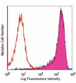
RAE-1δ transfected cells stained with Charlotte d1.23 PE
 Login / Register




Follow Us