- Clone
- A17129A (See other available formats)
- Regulatory Status
- RUO
- Other Names
- APG7, APG7-like, APG7L, Ubiquitin-activating enzyme E1-like protein, Ubiquitin-Like Modifier-Activating Enzyme ATG7, Autophagy-related protein 7
- Isotype
- Mouse IgG2b, κ
- Ave. Rating
- Submit a Review
- Product Citations
- publications
-

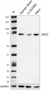
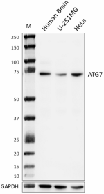
Whole tissue or cell extracts (15 µg total protein) were resolved by 4-12% Bis-Tris gel electrophoresis, transferred to a PVDF membrane, and probed with 1.0 µg/mL of purified anti-ATG7 (clone A17129A) overnight at 4°C. Proteins were visualized by chemiluminescence detection using HRP goat anti-mouse IgG (Cat. No. 405306) at a 1:3000 dilution. Direct-Blot™ HRP anti-GAPDH (Cat. No. 607903) was used as a loading control at a 1:50000 dilution. Western-Ready™ ECL Substrate Premium Kit (Cat. No. 426319) was used as a detection agent. Lane M: Molecular weight marker -

U-251 MG cells were fixed with Fixation Buffer (Cat. No. 420801) for 10 minutes and permeabilized with 0.5% Triton X-100 for 10 minutes, and blocked with 5% FBS for 1 hour at room temperature. Cells were then stained with 10 µg/mL of purified anti-ATG7 (clone A17129A), followed by incubation with Alexa Fluor® 647 Goat anti-mouse IgG (Cat. No. 405322) for 1 hour at room temperature (panel A). Cells were also stained with Flash Phalloidin™ Green 488 (Cat. No. 424201) for 1 hour at room temperature and nuclei were counterstained with DAPI (blue) (Cat. No. 422801) (merge in panel B). The images were captured with a 60X objective. Scale bar: 50 µm -

Rat primary neurons were fixed with Fixation Buffer (Cat. No. 420801) for 10 minutes and permeabilized with 0.5% Triton X-100 for 10 minutes, and blocked with 5% FBS for 1 hour at room temperature. Cells were then stained with 10 µg/mL of purified anti-ATG7 (clone A17129A), followed by incubation with Alexa Fluor® 647 goat anti-mouse IgG (Cat. No. 405322) for 1 hour at room temperature (panel A). Cells were also stained with Flash Phalloidin™ Green 488 (Cat. No. 424201) for 1 hour at room temperature and nuclei were counterstained with DAPI (blue) (Cat. No. 422801) (merge in panel B). The images were captured with a 60X objective. Scale bar: 50 µm -

IHC staining of purified anti-ATG7 (clone A17129A) on formalin-fixed paraffin-embedded human brain tissue. Following antigen retrieval using Citrate Buffer, 10X (Cat. No. 420902), the tissue was incubated with 5 µg/mL of the primary antibody overnight at 4°C. BioLegend’s Ultra Streptavidin HRP Kit (Multi-Species, DAB, Cat. No. 929501) was used for detection followed by hematoxylin counterstaining, according to the protocol provided. Images were captured with a 40X objective. Scale bar: 50 µm -

IHC staining of purified anti-ATG7 (clone A17129A) on formalin-fixed paraffin-embedded mouse brain tissue. Following antigen retrieval using Citrate Buffer, 10X (Cat. No. 420902), the tissue was incubated with 10 µg/mL of the primary antibody overnight at 4°C. BioLegend’s Ultra Streptavidin HRP Kit (Multi-Species, DAB, Cat. No. 929501) was used for detection followed by hematoxylin counterstaining, according to the protocol provided. Images were captured with a 40X objective. Scale bar: 50 µm -

IHC staining of purified anti-ATG7 (clone A17129A) on formalin-fixed paraffin-embedded rat brain tissue. Following antigen retrieval using Citrate Buffer, 10X (Cat. No. 420902), the tissue was incubated with 5 µg/mL of the primary antibody overnight at 4°C. BioLegend’s Ultra Streptavidin HRP Kit (Multi-Species, DAB, Cat. No. 929501) was used for detection followed by hematoxylin counterstaining, according to the protocol provided. Images were captured with a 40X objective. Scale bar: 50 µm
| Cat # | Size | Price | Quantity Check Availability | Save | ||
|---|---|---|---|---|---|---|
| 625851 | 25 µg | 136€ | ||||
| 625852 | 100 µg | 340€ | ||||
ATG7 is an E1-like ligase that plays a central role in autophagosome biogenesis by conjugating ATG5 to ATG12 which degrades and recycles damaged organelles, protein aggregates, and invading pathogens. ATG7 interacts with other ATG proteins, such as ATG3, ATG10, and ATG16L1, to form a complex that is required for autophagy, also regulates immunity, cell death, and protein secretion, and independently regulates the cell cycle and apoptosis. It also participates in lipidation of the ATG8 protein, which is essential for autophagosome formation. Additionally, ATG7 has been shown to interact with other proteins outside of the autophagy pathway, such as p53, to regulate apoptosis. Disrupting ATG7-dependent autophagy has been shown to cause electromotility disturbances, outer hair cell loss, and deafness in mice. ATG7 dysfunction has been associated with various human diseases, including cancer, neurodegeneration, and infection. ATG7 dysfunction and disease was found in patients with biallelic ATG7 variants and childhood-onset neuropathology. Furthermore, ATG7 has been shown to induce basal autophagy and rescue autophagic deficiency in CryABR120G cardiomyocytes.
Product DetailsProduct Details
- Verified Reactivity
- Human, Mouse, Rat
- Antibody Type
- Monoclonal
- Host Species
- Mouse
- Immunogen
- Recombinant fragment of human ATG7
- Formulation
- Phosphate-buffered solution, pH 7.2, containing 0.09% sodium azide
- Preparation
- The antibody was purified by affinity chromatography.
- Concentration
- 0.5 mg/mL
- Storage & Handling
- The antibody solution should be stored undiluted between 2°C and 8°C.
- Application
-
WB - Quality tested
ICC, IHC-P - Verified - Recommended Usage
-
Each lot of this antibody is quality control tested by western blotting. For western blotting, the suggested use of this reagent is 0.1 - 1.0 µg/mL. For immunocytochemistry, a concentration range of 5.0 - 10.0 μg/mL is recommended. For immunohistochemistry on formalin-fixed paraffin-embedded tissue sections, a concentration range of 5.0 - 10.0 µg/mL is suggested. It is recommended that the reagent be titrated for optimal performance for each application.
- Application Notes
-
For immunohistochemistry (IHC-P), Citrate Buffer, 10X (Cat. No, 420902) is recommended.
For immunocytochemistry (ICC), either Fixation Buffer (Cat. No. 420801) or paraformaldehyde-based fixation is recommended.
Clone A17129A detects homodimer of ATG7 in western blotting (WB). The homodimer band is demolished by increasing DTT concentration in the lysis buffer. - RRID
-
AB_3083415 (BioLegend Cat. No. 625851)
AB_3083415 (BioLegend Cat. No. 625852)
Antigen Details
- Structure
- Homodimer
- Distribution
-
Cytosol and preautophagosomal structure
- Function
- Conjugate ATG12 to ATG5, lipidation of ATG8
- Interaction
- Interacts with ATG3, ATG8, ATG12, EP300 and FOXO1
- Cell Type
- Mature Neurons, Neurons
- Biology Area
- Apoptosis/Tumor Suppressors/Cell Death, Cell Biology, Cell Death, Cell Proliferation and Viability, Neurodegeneration, Protein Misfolding and Aggregation, Protein Trafficking and Clearance, Ubiquitin/Protein Degradation
- Molecular Family
- Autophagosome Markers, Ligases
- Antigen References
-
- Komatsu M, et al. 2005. J Cell Biol. 169:425-34.
- Komatsu M, et al. 2007. Proc Natl Acad Sci USA. 104:14489-94.
- Lee I H, et al. 2012. Science. 336: 225–228.
- Collier JJ, et al. 2021. EMBO Mol Med. 13:e14824.
- Gene ID
- 10533 View all products for this Gene ID
- UniProt
- View information about ATG7 on UniProt.org
Related FAQs
Other Formats
View All ATG7 Reagents Request Custom Conjugation| Description | Clone | Applications |
|---|---|---|
| Purified anti-ATG7 | A17129A | WB,IHC-P,ICC |
Compare Data Across All Formats
This data display is provided for general comparisons between formats.
Your actual data may vary due to variations in samples, target cells, instruments and their settings, staining conditions, and other factors.
If you need assistance with selecting the best format contact our expert technical support team.
-
Purified anti-ATG7
Whole tissue or cell extracts (15 µg total protein) were res... 
U-251 MG cells were fixed with Fixation Buffer (Cat. No. 420... 
Rat primary neurons were fixed with Fixation Buffer (Cat. No... 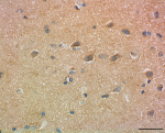
IHC staining of purified anti-ATG7 (clone A17129A) on formal... 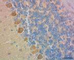
IHC staining of purified anti-ATG7 (clone A17129A) on formal... 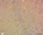
IHC staining of purified anti-ATG7 (clone A17129A) on formal...

 Login / Register


Follow Us