Purified anti-β-actin Antibody

Pricing & Availability
Clone
Poly6221 (See other available formats)
Regulatory Status
RUO
Other Names
Actin, cytoplasmic 1
Isotype
Rabbit Polyclonal IgG
Ave. Rating
Submit a Review
Product Citations
publications
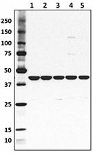
poly6221_PURE_betaactin_Antibody_1_WB_090616
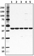
Total cell lysate from HeLa cells (lane 1, 15 µg), NIN3T3 cells (lane 2, 15 µg), U87-MG cells (lane 3, 15 µg), mouse brain (lane 4, 15 µg) and rat brain (lane 5, 15 µg) were resolved by electrophoresis (4-20% Tris-Glycine gel), transferred to nitrocellulose, and probed with purified anti-β-actin antibody (Poly6221). Proteins were visualized using an HRP Donkey anti-rabbit IgG Antibody and chemiluminescence detection.
  • poly6221_PURE_betaactin_Antibody_1_WB_090616
    Total cell lysate from HeLa cells (lane 1, 15 µg), NIN3T3 cells (lane 2, 15 µg), U87-MG cells (lane 3, 15 µg), mouse brain (lane 4, 15 µg) and rat brain (lane 5, 15 µg) were resolved by electrophoresis (4-20% Tris-Glycine gel), transferred to nitrocellulose, and probed with purified anti-β-actin antibody (Poly6221). Proteins were visualized using an HRP Donkey anti-rabbit IgG Antibody and chemiluminescence detection.
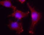
  • poly6221_PURE_betaactin_Antibody_2_IF_062016
    Immunofluorescent microscope analysis of Hela cells using Poly6221, followed by Rhodamine Red-X goat anti-rabbit IgG and DAPI.
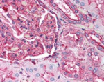
  • poly6221_PURE_betaactin_Antibody_3_IHCP_102720
    Formalin-fixed paraffin-embedded human kidney tissue was stained with Poly6221 and developed with an alkaline phosphatase chromogen substrate (red color). Tissue was counterstained with H&E (blue/pink). Magnification, 40X.
Cat # Size Price Quantity Check Availability Save
622101 50 µL 95€
Check Availability


Need larger quantities of this item?
Request Bulk Quote
622102 200 µL 235€
Check Availability


Need larger quantities of this item?
Request Bulk Quote
Description

β-actin is a ubiquitously expressed and highly conserved 42 kD cytoplasmic protein involved in cell motility. This critical cytoskeletal component can be disrupted by drugs such as cytochalasin. Because β-actin is ubiquitously expressed in all eukaryotic cells, it is frequently used as a loading control for assays involving protein detection (such as Western blotting). The Poly6221 antibody has been shown to be useful for Western blotting of mouse, rat, and human β-actin.

Product Details
Technical Data Sheet (pdf)

Product Details

Verified Reactivity
Mouse, Rat, Human
Antibody Type
Polyclonal
Host Species
Rabbit
Immunogen
Peptide-KLH, NH2 terminus
Formulation
This antibody is provided in phosphate-buffered solution, pH 7.2, containing 0.09% sodium azide and 50% glycerol.
Preparation
The antibody was purified by affinity chromatography.
Concentration
Lot-specific (to obtain lot-specific concentration and expiration, please enter the lot number in our Certificate of Analysis online tool.)
Storage & Handling
Upon receipt, store frozen at -20°C.
Application

WB - Quality tested
ICC, IHC-P - Verified

Recommended Usage

Each lot of this antibody is quality control tested by Western blotting. Western blotting, suggested working dilution(s): Use 10 µL per 5 mL antibody dilution buffer for each mini-gel. For immunocytochemistry: Use a dilution range of 1:100~1:400. For immunohistochemistry on formalin-fixed paraffin-embedded tissue sections, use a 1:100 dilution of antibody for staining. Antigen retrieval for IHC using 0.01 M sodium citrate buffer is recommended. It is recommended that the reagent be titrated for optimal performance for each application.

Application Notes

This product may contain other non-IgG subtypes.

Additional Product Notes
The 50 µL size can be used for approximately 5 Western blots, and the 200 µL size can be used for approximately 20 Western blots.
Application References
  1. Lawson BR, et al. 2007. J. Immunol. 178:5366.
  2. Joyce CW. 2006. J. Biol. Chem. 281:33053.
  3. Yanagiya T,et al.2007.Obesity.15:572.PubMed
  4. KishidaT,et al.2007.J. Immunol. 179:8554.PubMed
  5. Ouimet M, et al. 2008. Arterisocler Thromb Vasc Biol. 28:1144. PubMed
  6. Toltlt LJ,et al. 2008.J. Immunol. 181:2165. PubMed
  7. Sawada. T,et al. 2008. J. Biol. Chem. 283:26820. PubMed
  8. Ikeda D, et al. 2008. Endocrinology. 149:6037. PubMed
  9. Rahman MK, et al. 2010. J. Immunol. 184:7247. PubMed
  10. Shikama Y, et al. 2011. Innate Immun. 17:3. PubMed
  11. Zhang Y, et al. 2012. Am J Physiol Renal Physiol. 302:70. PubMed
  12. Piao X, et al. 2012. Sci Signal. 5:93. PubMed
  13. Ng CS, et al. 2013. J. Virol. 87:9511. PubMed
  14. Harvey BS, et al. 2014. Cytokine. 65:236. PubMed
  15. Diessner J, et al. 2014. Cell Death Dis. 5:1149. PubMed
  16. Hua X, et al. 2015. PloS One. 10:128039. PubMed
Product Citations
  1. Nishina T, et al. 2023. iScience. 26:105934. PubMed
  2. Checa M, et al. 2016. PLoS One. 11: 0150383. PubMed
  3. Okimoto T, et al. 2020. Cancer Sci. 111:1910. PubMed
  4. Al–Naggar IM, et al. 2019. J Gerontol A Biol Sci Med Sci. 74:325. PubMed
  5. Shibata K, et al. 2022. Nat Commun. 13:6948. PubMed
  6. Mete B, et al. 2022. Life Sci Alliance. 5:. PubMed
  7. Diessner J, et al. 2014. Cell Death Dis. 5:1149. PubMed
  8. Wang D, et al. 2018. Immunity. 48:659. PubMed
  9. Agabiti S, Li J, Wiemer A 2017. Cell Death Dis. 10.1038/cddis.2017.101. PubMed
  10. Wakatsuki S, et al. 2021. J Cell Biol. 220:. PubMed
  11. Piao X, et al. 2012. Sci Signal. 5:93. PubMed
  12. Kartika ID, et al. 2021. Cancer Med. 10:8988. PubMed
  13. Harvey B, et al. 2014. Cytokine. 65:236. PubMed
  14. Wakatsuki S, et al. 2015. J Cell Biol. 211: 881 - 896. PubMed
  15. Nishina T, et al. 2021. Nat Commun. 12:2281. PubMed
  16. Pfeil EM, et al. 2020. Molecular Cell. 80(6):940-954.e6. PubMed
  17. Hua X, et al. 2015. PLoS One. 10:128039. PubMed
  18. Che L, et al. 2019. Exp Ther Med. 18:1745. PubMed
  19. Ouimet M, et al. 2008. Arterioscler Thromb Vasc Biol. 28:1144. PubMed
  20. Rahman M, et al. 2010. J Immunol. 184:7247. PubMed
  21. Takeda K, et al. 2017. Nat Commun. 8:14607. PubMed
  22. Toltl L, et al. 2008. J Immunol. 181:2165. PubMed
  23. Tao HC, et al. 2020. Front Immunol. 11:148. PubMed
  24. Man F, et al. 2021. Am J Physiol Lung Cell Mol Physiol. 321:L803. PubMed
  25. Taha A, et al. 2020. J Cell Physiol. 7094:235. PubMed
  26. Nishigami Y, et al. 2016. Sci Rep. 6:18964. PubMed
  27. Berdasco C, et al. 2020. Front Cell Infect Microbiol. 0.681944444. PubMed
  28. Feddersen CR, et al. 2019. Cancer Res. 79:5074. PubMed
  29. Joyce C, et al. 2006. J Biol Chem. 281:33053. PubMed
  30. Zhu S et al. 2017. Cancer cell. 32(3):310-323 . PubMed
  31. Naneix F et al. 2017. eNeuro. 4(3) pii: ENEURO. PubMed
  32. Yanagiya T, et al. 2007. Obesity . 1.022222222. PubMed
  33. Kishida T, et al. 2007. J Immunol. 179:8554. PubMed
  34. Zheng X, et al. 2018. Int J Mol Med. 128:1300. PubMed
  35. Ikeda D, et al. 2008. Endocrinology. 149:6037. PubMed
  36. Caglar Beker M, et al. 2020. Turk J Med Sci. Online ahead of print. PubMed
  37. Zhang Y, et al. 2012. Am J Physiol Renal Physiol. 302:F70. PubMed
  38. Terrazas M 2014. Int J Parasit. 44 613 . PubMed
  39. Fliegauf M, et al. 2021. Front Immunol. 12:621503. PubMed
  40. Ng C, et al. 2013. J Virol. 87:9511. PubMed
  41. Grundmann M, et al. 2018. Nat Commun. 9:341. PubMed
  42. Sawada T, et al. 2008. J Biol Chem. 283:26820. PubMed
  43. Pototskiy E, et al. 2021. Sci Rep. 11:17262. PubMed
  44. Tsurusaki S, et al. 2019. Cell Death Dis. 10:449. PubMed
  45. Panwar P, et al. 2018. Br J Pharmacol. 175:902. PubMed
  46. Merten N et al. 2018. Cell chemical biology. 25(6):775-786 . PubMed
  47. Kumarasinghe N, et al. 2019. BMC Res Notes. 12:33. PubMed
  48. Inao T, et al. 2019. Cancer Sci. 110:2690. PubMed
  49. Shikama Y, et al. 2011. Innate Immunity. 17:03. PubMed
  50. Ergünay T, et al. 2022. Life Sci Alliance. 5:. PubMed
RRID
SCR_001134 (BioLegend Cat. No. 622101)
SCR_001134 (BioLegend Cat. No. 622102)

Antigen Details

Structure
Member of the actin family; 42 KD
Distribution

Ubiquitously expressed in the cytoplasm of all eukaryotic cells

Function
Actins are highly conserved proteins that are involved in cell motility
Interaction
Polymerization of globular actin (G-actin) leads to a structural filament (F-actin) that forms a two-stranded helix. Each actin can bind to four others.
Biology Area
Cell Biology, Cell Motility/Cytoskeleton/Structure, Neuroscience, Neuroscience Cell Markers
Molecular Family
Microfilaments
Antigen References

1. Hanukoglu I, et al. 1983. J. Mol. Biol. 163:673.
2. Nakajima-Iijima S, et al. 1985. Proc. Natl. Acad. Sci. 82:6133.
3. Ponte P, et al. 1984. Nucleic Acids Res. 12:1687.

Regulation
Three actin isoforms exist in vertebrates; alpha, found in muscle tissues; beta and gamma, found as cytoskeletal components and mediators of internal cell motility. Drugs such as cytochalasin disrupt the actin network.
Gene ID
11461 View all products for this Gene ID
UniProt
View information about beta-actin on UniProt.org

Related FAQs

There are no FAQs for this product.
Go To Top Version: 6    Revision Date: 10-27-2020

For Research Use Only. Not for diagnostic or therapeutic use.


This product is supplied subject to the terms and conditions, including the limited license, located at www.biolegend.com/terms) ("Terms") and may be used only as provided in the Terms. Without limiting the foregoing, BioLegend products may not be used for any Commercial Purpose as defined in the Terms, resold in any form, used in manufacturing, or reverse engineered, sequenced, or otherwise studied or used to learn its design or composition without express written approval of BioLegend. Regardless of the information given in this document, user is solely responsible for determining any license requirements necessary for user’s intended use and assumes all risk and liability arising from use of the product. BioLegend is not responsible for patent infringement or any other risks or liabilities whatsoever resulting from the use of its products.


BioLegend, the BioLegend logo, and all other trademarks are property of BioLegend, Inc. or their respective owners, and all rights are reserved.


8999 BioLegend Way, San Diego, CA 92121 www.biolegend.com
Toll-Free Phone: 1-877-Bio-Legend (246-5343) Phone: (858) 768-5800 Fax: (877) 455-9587

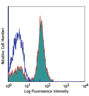
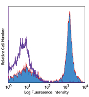
This data display is provided for general comparisons between formats.
Your actual data may vary due to variations in samples, target cells, instruments and their settings, staining conditions, and other factors.
If you need assistance with selecting the best format contact our expert technical support team.

ProductsHere

Login / Register
Remember me
Forgot your password? Reset password?
Create an Account