
 Login / Register


- Clone
- 7H8.2C12 (See other available formats)
- Regulatory Status
- RUO
- Other Names
- Cyt c
- Isotype
- Mouse IgG2b, κ
- Ave. Rating
- Submit a Review
- Product Citations
- 9 publications


| Cat # | Size | Price | Quantity Check Availability | Save | ||
|---|---|---|---|---|---|---|
| 612503 | 50 µL | 85€ | ||||
| 612504 | 200 µL | 235€ | ||||
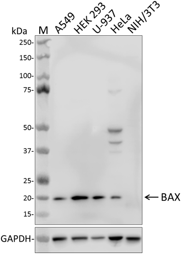
Cytochrome c is a 15 kD protein found in the mitochondrial intermembrane space with a heme-binding domain. Cytochrome c is a component of the electron transport chain; the heme group transfers electrons from cytochrome b-c1 complex to cytochrome oxidase complex. Cytochrome c initiates apoptosis by release to cytoplasm and binding Apaf-1 which activates procaspase 9. Cytochrome c interacts with the cytochrome b-c1 complex, cytochrome oxidase complex, heme, Apaf-1, and Caspase 9 proteins. The 7H8.2C12 monoclonal antibody recognizes cytochrome-c from most species and has been shown to be useful for Western blotting.
Product DetailsProduct Details
- Verified Reactivity
- Human, Mouse, Rat, Hamster
- Reported Reactivity
- Other species
- Antibody Type
- Monoclonal
- Host Species
- Mouse
- Immunogen
- Horse cyt c-OVA
- Formulation
- This antibody is provided in phosphate-buffered solution, pH 7.2, containing 0.09% sodium azide.
- Preparation
- The antibody was purified by affinity chromatography.
- Concentration
- Lot-specific (to obtain lot-specific concentration and expiration, please enter the lot number in our Certificate of Analysis online tool.)
- Storage & Handling
- Upon receipt, store undiluted between 2°C and 8°C.
- Application
-
WB - Quality tested
ICC - Verified - Recommended Usage
-
Each lot of this antibody is quality control tested by Western blotting. Western blotting, suggested working dilution(s): Use 10 µl antibody per 5 ml antibody dilution buffer for each mini-gel. For immunocytochemistry, a concentration range of 1.0 - 3.0 µg/ml (1:150-1:500 dilution) is recommended. It is recommended that the reagent be titrated for optimal performance for each application.
- Additional Product Notes
- The 50 µL size can be used for approximately 5 Western blots, and the 200 µL size can be used for approximately 20 Western blots.
- Application References
-
- Jemmerson R, et al. 1991. Eur. J. Immunol. 21:143. (WB)
- Semenkova L, et al. 2003. Eur. J. Biochem. 270:4388. (WB)
- Product Citations
-
- RRID
-
AB_2090157 (BioLegend Cat. No. 612503)
AB_2292697 (BioLegend Cat. No. 612504)
Antigen Details
- Structure
- Heme binding domain; 15 kD
- Distribution
-
Mitochondrial intermembrane space
- Function
- Component of electron transport chain; heme group transfers electrons from cytochrome b-c1 complex to cytochrome oxidase complex. Initiates apoptosis by release to cytoplasm and binding Apaf-1 which activates procaspase 9
- Interaction
- Cytochrome b-c1 complex, cytochrome oxidase complex, heme, Apaf-1, Casp9
- Biology Area
- Apoptosis/Tumor Suppressors/Cell Death, Cell Biology, Mitochondrial Function, Neuroscience, Neuroscience Cell Markers
- Molecular Family
- Mitochondrial Markers
- Antigen References
-
1. Liu X, et al. 1996. Cell 86:147.
2. Li P, et al. 1997. Cell 91:479.
3. Zhang Z, et al. 2003. Gene 312:61.
4. Ferguson H, et al. 2003. J. Biol. Chem. 278:45793. - Gene ID
- 54205 View all products for this Gene ID
- UniProt
- View information about Cytochrome c on UniProt.org

Related FAQs
Other Formats
View All Cytochrome c Reagents Request Custom Conjugation| Description | Clone | Applications |
|---|---|---|
| Purified anti-Cytochrome c | 7H8.2C12 | WB,ICC |
Customers Also Purchased

Compare Data Across All Formats
This data display is provided for general comparisons between formats.
Your actual data may vary due to variations in samples, target cells, instruments and their settings, staining conditions, and other factors.
If you need assistance with selecting the best format contact our expert technical support team.


Follow Us