- Clone
- W19410A (See other available formats)
- Regulatory Status
- RUO
- Other Names
- Eukaryotic Translation Elongation Factor 2, Polypeptidyl-tRNA Translocase, Epididymis Secretory Sperm Binding Protein
- Isotype
- Rat IgG2b, κ
- Ave. Rating
- Submit a Review
- Product Citations
- publications

-

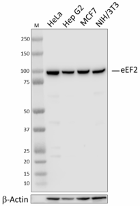
Total cell lysates (15 µg total protein) from the indicated cell lines were resolved by 4-12% Bis-Tris gel electrophoresis, transferred to a PVDF membrane, and probed with a 1:8000 dilution of purified anti-eEF2 antibody (clone W19410A) overnight at 4°C. Proteins were visualized by chemiluminescence detection using HRP goat anti-rat IgG antibody (Cat. No. 405405) at a 1:3000 dilution. Direct-Blot™ HRP anti-β-actin antibody (Cat. No. 664803) was used as a loading control at a 1:25000 dilution (lower). Western-Ready™ ECL Substrate Premium Kit (Cat. No. 426319) was used as a detection agent. Lane M: Molecular weight marker. -

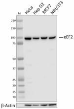
ICC staining of purified anti-eEF2 antibody (clone W19410A) on eEF2-depleted HeLa cells. HeLa cells treated with control siRNA (panel A) or siRNA targeting eEF2 (panel B) were fixed and permeabilized with 100% methanol and blocked with 5% FBS for 1 hour. The cells were then stained with a 1:100 dilution of the primary antibody, followed by incubation with 2.5 µg/mL of Alexa Fluor® 594 anti-goat anti-rat IgG antibody (Cat. No. 405422) for 1 hour at room temperature. Nuclei were counterstained with DAPI, and the images were captured with a 60X objective. -

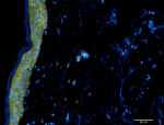
IHC staining of purified anti-eEF2 antibody (clone W19410A) on formalin-fixed paraffin-embedded human skin tissue. The tissue was incubated with a 1:100 dilution of the primary antibody overnight at 4°C, followed by incubation with 2.5 μg/mL of Alexa Fluor® 647 goat anti-mouse IgG (yellow) (Cat. No. 405322) for one hour at room temperature. Nuclei were counterstained with DAPI (blue), and the slide was mounted with ProLong™ Gold Antifade Mountant. The image was captured with a 10x objective. Scale bar = 50 μm. -

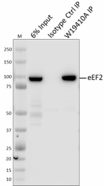
Whole cell extracts (250 µg total protein) prepared from HeLa cells were immunoprecipitated overnight with 2.5 µg of purified rat IgG2b, κ Isotype ctrl antibody (Cat. No. 400602) or 25 µL of purified anti-eEF2 antibody (clone W19410A). The resulting IP fractions and whole cell extract input (6%) were resolved by 4-12% Bis-Tris gel electrophoresis, transferred to a PVDF membrane, and probed with a control antibody against a separate epitope of eEF2. Lane M: Molecular weight marker.
| Cat # | Size | Price | Quantity Check Availability | Save | ||
|---|---|---|---|---|---|---|
| 947601 | 25 µL | 95€ | ||||
| 947602 | 100 µL | 235€ | ||||
eEF2 is a eukaryotic elongation factor required for GTP-dependent ribosomal translocation and is negatively regulated by eEF2 kinase. In response to elevated cytosolic calcium, calmodulin binds to and stimulates eEF2 kinase activity, stimulating phosphorylation and inactivation of eEF2 elongation function. Hippocampal tissue from Alzheimer’s patients display hyperphosphorylation of eEF2 at threonine 56 compared to age-matched controls. Emerging studies also show that sub-psychomimetic doses of ketamine, which is used as a rapid-acting anti-depressant, result in a block of NMDAR and deactivation of eEF2 kinase, leading to reduced phosphorylation of eEF2 and increased eEF2-dependent translation of brain-derived neurotrophic factor.
Product DetailsProduct Details
- Verified Reactivity
- Human, Mouse
- Antibody Type
- Monoclonal
- Host Species
- Rat
- Immunogen
- Partial recombinant human eEF2 protein
- Formulation
- Phosphate buffer, 0.5% BSA, 0.09% NaN3, bottle antibody at 0.1 mg/mL
- Preparation
- The antibody was purified by affinity chromatography.
- Concentration
- 0.1 mg/mL
- Storage & Handling
- The antibody solution should be stored undiluted between 2°C and 8°C.
- Application
-
WB - Quality tested
ICC, IHC-P, IP, KO/KD-WB - Verified - Recommended Usage
-
Each lot of this antibody is quality control tested by western blotting. For western blotting, the suggested use of this reagent is a 1:8000 dilution. For immunocytochemistry, a dilution of 1:100 dilution is recommended. For immunohistochemistry, a dilution of 1:100 dilution is suggested. For immunoprecipitation, the suggested use of this reagent is 25 µg/test. It is recommended that the reagent be titrated for optimal performance for each application.
- Application Notes
-
When used for western blotting, W19410A antibody concentrations exceeding 0.0125 µg/mL may produce non-specific binding.
Clone W9410A was tested for ICC using the following fixation-permeabilization methods: 4% PFA + Triton X-100, 4% PFA + methanol, and methanol-only. All of these methods were compatible with eEF2 staining.
BioLegend offers two sizes (25 and 100 µL0 for W19410A. If using this clone for immunoprecipitation, we recommend purchasing the 100 µL size to ensure sufficient material for the application. - RRID
-
AB_2904459 (BioLegend Cat. No. 947601)
AB_2904459 (BioLegend Cat. No. 947602)
Antigen Details
- Structure
- eEF2 is an 858 amino acid protein with a predicted molecular weight of 95 kD.
- Distribution
-
Ubiquitously expressed/Cytosol
- Function
- Protein translation elongation factor
- Biology Area
- Cell Biology, Protein Synthesis
- Antigen References
-
1. Ryazanov AG, et al. 1991. FEBS. Lett. 285: 170.
2. Autry AE, et al. 2011. Nature. 475: 91.
3. Beckelman BC, et al. 2019. J. Clin. Invest. 129: 820. - Gene ID
- 1938 View all products for this Gene ID
- UniProt
- View information about eEF2 on UniProt.org
Related Pages & Pathways
Pages
Related FAQs
Other Formats
View All eEF2 Reagents Request Custom Conjugation| Description | Clone | Applications |
|---|---|---|
| Purified anti-eEF2 Antibody | W19410A | WB,IHC-P,ICC,IP,KO/KD-WB |
Compare Data Across All Formats
This data display is provided for general comparisons between formats.
Your actual data may vary due to variations in samples, target cells, instruments and their settings, staining conditions, and other factors.
If you need assistance with selecting the best format contact our expert technical support team.

 Login / Register


Follow Us