- Clone
- W16161B (See other available formats)
- Regulatory Status
- RUO
- Other Names
- Early Growth Response 1, ZNF225, NGFI-A, TIS8, AT225, G0S30, KROX-24, ZIF-268, Transcription Factor ETR103
- Isotype
- Rat IgG2a, κ
- Ave. Rating
- Submit a Review
- Product Citations
- publications
-

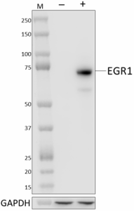
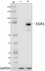
Whole cell extracts (15 µg total protein) from serum-starved HeLa cells untreated (-) or treated with 100 ng/mL recombinant human EGF (+) (Cat. No. 585506) for 1 hour were resolved by 4-12% Bis-Tris gel electrophoresis, transferred to a PVDF membrane, and probed with 1.0 µg/mL (1:500 dilution) purified anti-EGR1 antibody (clone W16161B) overnight at 4°C. Proteins were visualized by chemiluminescence detection using HRP goat anti-rat IgG antibody (Cat. No. 405405) at a 1:3000 dilution. Direct-Blot™ HRP anti-GAPDH antibody (Cat. No. 607904) was used as a loading control at a 1:25000 dilution (lower). Lane M: Molecular weight marker. -

Serum-starved untreated HeLa cells (panel A) and HeLa cells treated with 100 ng/mL recombinant human EGF (Cat. No. 585506) for 1 hour (panel B) were fixed with 4% paraformaldehyde for 10 minutes, permeabilized with Triton X-100 for 10 minutes, and blocked with 5% FBS for 60 minutes. Cells were then intracellularly stained with purified anti-EGR1 antibody (clone W16161B) overnight at 4°C followed by incubation with Alexa Fluor® 594 goat anti-rat IgG (Cat. No. 405422) at 2.0 µg/mL. Nuclei were counterstained with DAPI and the image was captured with a 60X objective. -

Whole cell extracts (250 µg total protein) prepared from HeLa cells treated with 100 ng/mL EGF for 1 hour were immunoprecipitated overnight with 2.0 µg of purified rat IgG2a, κ isotype ctrl antibody (Cat. No. 400501) or purified anti-EGR1 antibody (clone W16161B). The resulting IP fractions and whole cell extract input (6%) were resolved by 4-12% Bis-Tris gel electrophoresis, transferred to a PVDF membrane and probed with a rabbit control antibody against a separate epitope of EGR1. Lane M: Molecular weight marker. -

Whole cell extracts (15 µg total protein) from serum-starved NIH/3T3 cells untreated (-) or treated with 100 ng/mL recombinant human EGF (+) (Cat. No. 585506) for 1 hour were resolved by 4-12% Bis-Tris gel electrophoresis, transferred to a PVDF membrane, and probed with 1.0 µg/mL (1:500 dilution) purified anti-EGR1 antibody (clone W16161B) overnight at 4°C. Proteins were visualized by chemiluminescence detection using HRP goat anti-rat IgG antibody (Cat. No. 405405) at a 1:3000 dilution. Direct-Blot™ HRP anti-GAPDH antibody (Cat. No. 607904) was used as a loading control at a 1:25000 dilution (lower). Lane M: Molecular weight marker. -

HeLa cells treated (filled histogram, positive control) or untreated (open histogram, negative control) with 100 ng of recombinant human EGF (Cat. No. 585508) for 1 hour were fixed and permeabilized using the True-Phos™ Perm Buffer set (Cat. No. 420801, 425401), and intracellularly stained with purified anti-EGR1 antibody (clone W16161B) followed by PE goat anti-rat IgG antibody (Cat. No. 405406).
| Cat # | Size | Price | Quantity Check Availability | Save | ||
|---|---|---|---|---|---|---|
| 943901 | 25 µg | 95€ | ||||
| 943902 | 100 µg | 235€ | ||||
EGR1 (early growth response protein 1) is a zinc transcription factor vital to numerous physiological processes including cell cycle, differentiation, proliferation, metabolism, DNA damage response, and apoptosis. It also functions in mitogenesis, tissue repair, fibrosis, inflammatory response and immune response through its regulation of neutrophil gene expression and association with immune response genes such as TNFα, VEGF, and matrix metalloproteinases. In the central nervous system, EGR1 functions as an immediate early gene (IEG) and is vital to neuronal function and signaling, synaptic plasticity, cognition, and memory formation. Activated by acute mechanical injury and vascular stress, EGR1 regulates multiple cardiovascular pathological processes including atherosclerosis, intimal thickening and vascular repair in response to vascular injury, ischemia, angiogenesis, and cardiac hypertrophy. EGR1 is essential to the development, homeostasis, and repair of connective tissues through its regulation of genes of the extracellular matrix. EGR1 is a component of some oncogenic pathways and stimulates tumor growth and proliferation in prostate and gastric cancers. It also regulates a variety of tumor suppressors such as p53, PTEN, NAG-1, and TGFβ1, contributes to DNA repair, and induces apoptosis of tumor cells, thus functioning in dual roles as both an oncogene and as a tumor suppressor.
Product DetailsProduct Details
- Verified Reactivity
- Human, Mouse
- Antibody Type
- Monoclonal
- Host Species
- Rat
- Immunogen
- Partial recombinant human EGR1 protein
- Formulation
- Phosphate-buffered solution, pH 7.2, containing 0.09% sodium azide
- Preparation
- The antibody was purified by affinity chromatography.
- Concentration
- 0.5 mg/mL
- Storage & Handling
- The antibody solution should be stored undiluted between 2°C and 8°C.
- Application
-
WB - Quality tested
ICC, IP, ICFC - Verified - Recommended Usage
-
Each lot of this antibody is quality control tested by western blotting. For western blotting, the suggested use of this reagent is 0.125 - 1.0 µg/mL. For immunocytochemistry, a concentration range of 5.0 - 10 μg/mL is recommended. For immunoprecipitation, the suggested use of this reagent is 2.0 µg/test. For flow cytometric staining, the suggested use of this reagent is ≤ 0.5 µg per million cells in 100 µL volume. It is recommended that the reagent be titrated for optimal performance for each application.
- Application Notes
-
When using this clone for ICFC, we recommend using the True Phos perm buffer. We do not recommend using the True-Nuclear™ Transcription Factor Buffer Set or the Intracellular Staining Permeabilization Wash Buffer for ICFC testing due to poor EGR1 staining.
- RRID
-
AB_2890867 (BioLegend Cat. No. 943901)
AB_2890867 (BioLegend Cat. No. 943902)
Antigen Details
- Structure
- EGR1 is a 533 amino acid protein with a predicted molecular weight of 57 kD. The observed molecular weight ranges from 75 – 100 kD due to post-translational modifications.
- Distribution
-
Ubiquitously expressed/ Nucleus and cytoplasm
- Function
- Transcription factor
- Cell Type
- Neurons
- Biology Area
- Cell Biology, Transcription Factors
- Antigen References
-
- Cullen EM, et al. 2010. Mol Immunol. 47:1701-9.
- Duclot F & Kabbaj M. 2017. Front Behav Neurosci. 11:35.
- Havis E & Duprez D. 2020. Int J Mol Sci. 21:1664.
- Khachigian LM. 2006. Circ Res. 98:186-91.
- Li T, et al. 2019. Med Oncol. 37:7.
- Gene ID
- 1958 View all products for this Gene ID
- UniProt
- View information about EGR1 on UniProt.org
Related FAQs
Other Formats
View All EGR1 Reagents Request Custom Conjugation| Description | Clone | Applications |
|---|---|---|
| Purified anti-EGR1 | W16161B | WB,ICC,IP,ICFC |
| Alexa Fluor® 647 anti-EGR1 | W16161B | ICFC,ICC |
Compare Data Across All Formats
This data display is provided for general comparisons between formats.
Your actual data may vary due to variations in samples, target cells, instruments and their settings, staining conditions, and other factors.
If you need assistance with selecting the best format contact our expert technical support team.
-
Purified anti-EGR1
Whole cell extracts (15 µg total protein) from serum-starved... 
Serum-starved untreated HeLa cells (panel A) and HeLa cells ... 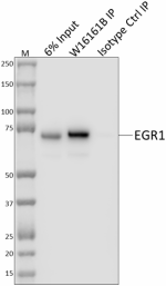
Whole cell extracts (250 µg total protein) prepared from HeL... 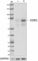
Whole cell extracts (15 µg total protein) from serum-starved... 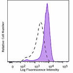
HeLa cells treated (filled histogram, positive control) or u... -
Alexa Fluor® 647 anti-EGR1
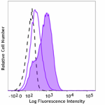
HeLa cells treated with 100 ng/mL recombinant human EGF (Cat... 
ICC staining of Alexa Fluor® 647 anti-EGR1 (clone W16161B) o...

 Login / Register


Follow Us