- Clone
- HM47 (See other available formats)
- Regulatory Status
- RUO
- Workshop
- V cB017
- Other Names
- Mb-1, Iga, CD79
- Isotype
- Mouse IgG1, κ
- Ave. Rating
- Submit a Review
| Cat # | Size | Price | Quantity Check Availability | Save | ||
|---|---|---|---|---|---|---|
| 333502 | 100 µg | 92€ | ||||
CD79a is a 47 kD type I integral membrane protein, also known as mb-1 or Iga. It is a member of the Ig superfamily and disulphide-associated with CD79b (B29). The interaction of CD79a/CD79b heterodimer with B cell suface Ig forms B cell antigen receptor complex. CD79a is expressed in B cells from early pre-B to plasma cell stage. It has been shown that CD79a is also weakly expressed in some precursors of T- and myeloid cells. CD79 mediates the transport of IgM to B cell surface and transduces signals initiated by BCR aggregation.
Product DetailsProduct Details
- Verified Reactivity
- Human
- Antibody Type
- Monoclonal
- Host Species
- Mouse
- Formulation
- Phosphate-buffered solution, pH 7.2, containing 0.09% sodium azide.
- Preparation
- The antibody was purified by affinity chromatography.
- Concentration
- 0.5 mg/ml
- Storage & Handling
- The antibody solution should be stored undiluted between 2°C and 8°C.
- Application
-
ICFC - Quality tested
IHC-P - Verified
IP, WB - Reported in the literature, not verified in house - Recommended Usage
-
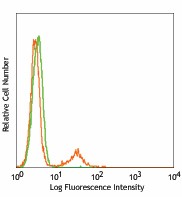
Each lot of this antibody is quality control tested by intracellular immunofluorescent staining with flow cytometric analysis. For flow cytometric staining, the suggested use of this reagent is ≤ 0.125 µg per million cells in 100 µl volume. For immunohistochemical staining on formalin-fixed paraffin-embedded tissue sections, the suggested use of this reagent is 5 - 10 µg per ml. It is recommended that the reagent be titrated for optimal performance for each application.
- RRID
-
AB_1089078 (BioLegend Cat. No. 333502)
Antigen Details
- Structure
- 47 kD, type I integral protein, Ig superfamily, CD79a/CD79b heterodimer interacts with B cell membrane Ig.
- Distribution
-
B cells (from early pre-B to plasma cell stage), some T and myeloid cell precursors
- Function
- In association with CD79b and B cell membrane Ig to form B cell antigen receptor complex
- Cell Type
- B cells, Hematopoietic stem and progenitors
- Biology Area
- Immunology
- Molecular Family
- CD Molecules
- Antigen References
-
1. Zola Heddy, et al. Eds. 2007. Leukocyte and Stromal Cell Molecules:The CD markers. WILEY-LISS
2. Astsaturov IA, et al. 1996. Leukemia 10:769
3. Mson DY, et al. 1995 Blood 86:1453
4. Hashimoto M, et al. 2002. J. Pathol. 197:341 - Gene ID
- 973 View all products for this Gene ID
- UniProt
- View information about CD79a on UniProt.org
Related FAQs
Other Formats
View All Reagents Request Custom Conjugation| Description | Clone | Applications |
|---|---|---|
| Purified anti-human CD79a (Igα) | HM47 | ICFC,IHC-P,IP,WB |
| PE anti-human CD79a (Igα) | HM47 | ICFC,SB |
| APC anti-human CD79a (Igα) | HM47 | ICFC |
| PerCP/Cyanine5.5 anti-human CD79a (Igα) | HM47 | ICFC |
| PE/Cyanine7 anti-human CD79a (Igα) | HM47 | ICFC |
| FITC anti-human CD79a (Igα) | HM47 | ICFC |
| Alexa Fluor® 594 anti-human CD79a (Igα) | HM47 | IHC-P |
| Alexa Fluor® 647 anti-human CD79a (Igα) | HM47 | ICFC,IHC-P |
| APC anti-human CD79a | HM47 | ICFC |
| PerCP/Cyanine5.5 anti-human CD79a | HM47 | ICFC |
| PE anti-human CD79a | HM47 | ICFC |
| FITC anti-human CD79a | HM47 | ICFC |
| TotalSeq™-B0578 anti-human CD79a (Igα) | HM47 | ICPG |
| GMP PerCP/Cyanine5.5 anti-human CD79a (Igα) | HM47 | ICFC |
| Brilliant Violet 421™ anti-human CD79a (Igα) | HM47 | ICFC |
| GMP APC anti-human CD79a (Igα) | HM47 | ICFC |
| TotalSeq™-C0578 anti-human CD79a (Igα) Antibody | HM47 | ICPG |
Customers Also Purchased
Compare Data Across All Formats
This data display is provided for general comparisons between formats.
Your actual data may vary due to variations in samples, target cells, instruments and their settings, staining conditions, and other factors.
If you need assistance with selecting the best format contact our expert technical support team.
-
Purified anti-human CD79a (Igα)
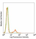
Human peripheral blood lymphocytes intracellularly stained w... 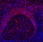
Human paraffin-embedded tonsil tissue slices were prepared w... -
PE anti-human CD79a (Igα)
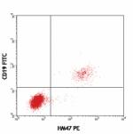
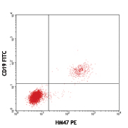
Human peripheral blood lymphocytes surface stained with CD19... 
Multiplexed IHC staining of PE anti-CD79a (clone HM47) on fo... -
APC anti-human CD79a (Igα)
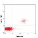
Human peripheral blood lymphocytes surface stained with CD19... -
PerCP/Cyanine5.5 anti-human CD79a (Igα)

Human peripheral blood lymphocytes surface stained with CD19... -
PE/Cyanine7 anti-human CD79a (Igα)

Human peripheral blood lymphocytes were stained with CD19 Br... -
FITC anti-human CD79a (Igα)
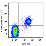
Human peripheral blood lymphocytes were stained with CD19 AP... 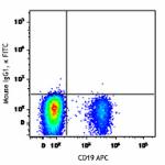
-
Alexa Fluor® 594 anti-human CD79a (Igα)
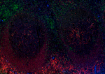
Human paraffin-embedded tonsil tissue slices were prepared w... -
Alexa Fluor® 647 anti-human CD79a (Igα)

Human peripheral blood lymphocytes surface stained with CD19... 
Human paraffin-embedded tonsil tissue slices were prepared w... -
APC anti-human CD79a
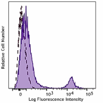
Typical results from human peripheral blood lymphocytes were... -
PerCP/Cyanine5.5 anti-human CD79a
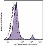
Typical results from human peripheral blood lymphocytes fixe... -
PE anti-human CD79a
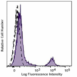
Typical results from human peripheral blood lymphocytes were... -
FITC anti-human CD79a
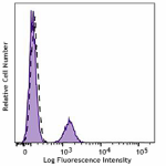
Typical results from human peripheral blood lymphocytes fixe... -
TotalSeq™-B0578 anti-human CD79a (Igα)
-
GMP PerCP/Cyanine5.5 anti-human CD79a (Igα)

Typical results from human peripheral blood lymphocytes fixe... -
Brilliant Violet 421™ anti-human CD79a (Igα)

Human peripheral blood lymphocytes were surface stained with... -
GMP APC anti-human CD79a (Igα)

Typical results from human peripheral blood lymphocytes were... -
TotalSeq™-C0578 anti-human CD79a (Igα) Antibody

 Login / Register




Follow Us