- Clone
- W19167A (See other available formats)
- Regulatory Status
- RUO
- Other Names
- Paired Box 6, Paired Box Protein PAX-6, Aniridia type II protein, Oculorhombin, AN2, D11S812E, MGDA, AN, MGC17209, WAGR, Aniridia 1, Aniridia 2, ASGD5, FVH1
- Isotype
- Rat IgG2a, κ
- Ave. Rating
- Submit a Review
- Product Citations
- publications

-

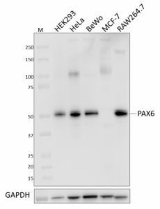
Whole cell extracts (15 µg total protein) from the indicated cell lines were resolved by 4-12% Bis-Tris gel electrophoresis, transferred to a PVDF membrane, and probed with 0.25 µg/mL (1:2000 dilution) purified anti-PAX6 antibody (clone W19167A) overnight at 4°C. Proteins were visualized by chemiluminescence detection using HRP goat anti-rat IgG antibody (Cat. No. 405405) at a 1:3000 dilution. Direct-Blot™ HRP anti-GAPDH antibody (Cat. No. 607904) was used as a loading control at a 1:25000 dilution (lower). Lane M: Molecular weight marker. -

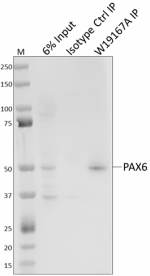
Whole cell extracts (250 µg total protein) prepared from HeLa cells were immunoprecipitated overnight with 2.0 µg of purified rat IgG2a, κ isotype ctrl antibody (Cat. No. 400502) or purified anti-PAX6 antibody (clone W19167A). The resulting IP fractions and whole cell extract input (6%) were resolved by 4-12% Bis-Tris gel electrophoresis, transferred to a PVDF membrane and probed with purified anti-PAX6 antibody (clone AD2.35) (Cat. No. 862002). Lane M: Molecular weight marker. -

IHC staining of purified anti-PAX6 antibody (clone W19167A) on frozen cerebellum brain (A, positive control) and sciatic nerve (B, negative control) tissues. Following fixation (Cat. No. 420801) and permeabilization with 0.5% Triton X-100, the tissue sections were incubated with 0.5 µg/mL of purified anti-PAX6 antibody overnight at 4°C. Alexa Fluor® 594 anti-rat IgG2a antibody (Cat. No. 407509) was used for 2-hour incubation at room temperature, followed by nuclei counterstain with DAPI (Cat. No. 422801). The slides were then mounted with ProLong™ Gold Antifade Mountant for image acquisition with a 40X objective. Scale bar: 50 µm.
| Cat # | Size | Price | Quantity Check Availability | Save | ||
|---|---|---|---|---|---|---|
| 939801 | 25 µg | 95€ | ||||
| 939802 | 100 µg | 235€ | ||||
Paired box protein 6 (PAX6) is a transcription factor with dual roles as both transcriptional activator and repressor. It plays a critical role in the regulation of cell proliferation, cell cycle length and exit, and cell differentiation during neurogenesis and ocular, cortical, and olfactory development. PAX6 forms a complex with SOX2 and δ-crystallin enhancer DC5 to initiate lens placode development. In the central nervous system, PAX6 functions in the regulation of progenitor cell proliferation, telencephalic patterning, cortical layer formation, and the maintenance of balance in neuron differentiation. Heterozygous mutations in PAX6 can result in aniridia, nystagmus, neurodevelopmental disorders, and auditory and memory impairment. In the neuroendocrine system, PAX6 functions in the development of β cells, pancreatic islet of Langerhans cells, and the pituitary gland as well as in insulin biosynthesis, and glucose-induced insulin secretion. PAX6 functions as a tumor suppressor in prostate cancer and glioblastoma by inhibiting androgen receptors.
Product DetailsProduct Details
- Verified Reactivity
- Human, Mouse
- Antibody Type
- Monoclonal
- Host Species
- Rat
- Immunogen
- Synthetic peptide corresponding to C-terminus of human PAX6
- Formulation
- Phosphate-buffered solution, pH 7.2, containing 0.09% sodium azide
- Preparation
- The antibody was purified by affinity chromatography.
- Concentration
- 0.5 mg/mL
- Storage & Handling
- The antibody solution should be stored undiluted between 2°C and 8°C.
- Application
-
WB - Quality tested
IP, IHC-F - Verified - Recommended Usage
-
Each lot of this antibody is quality control tested by western blotting. For western blotting, the suggested use of this reagent is 0.05 - 0.25 µg/mL. For immunoprecipitation, the suggested use of this reagent is 2.0 µg/test. For immunohistochemistry on frozen tissue sections, a concentration range of 0.5 - 5.0 µg/mL is suggested. It is recommended that the reagent be titrated for optimal performance for each application.
- Application Notes
-
At higher concentrations, this clone recognizes a ~100 kD protein of unknown origin. This protein was not detected in W19167A immunoprecipitates that were probed by western blot using W19167A.
- RRID
-
AB_2888904 (BioLegend Cat. No. 939801)
AB_2888904 (BioLegend Cat. No. 939802)
Antigen Details
- Structure
- PAX6 is a 422 amino acid protein with a predicted molecular weight of 47 kD.
- Distribution
-
Expressed in the eye, nose, brain pancreas/Nucleoplasm
- Function
- Transcriptional regulation, differentiation
- Antigen References
-
1. Gosmain Y, B, et al. 2012. Mol.. Endocrinol. 26:696
2. Kamachi Y, et al. 2001. Genes Dev. 10:1272
3. Manuel M, et al. 2015. Fron. Cell. Neurosci. 9:70
4. Shyr C, et al. 2010. Prostate. 274:6056 - Gene ID
- 5080 View all products for this Gene ID
- UniProt
- View information about PAX6 on UniProt.org
Related Pages & Pathways
Pages
Related FAQs
Other Formats
View All Reagents Request Custom Conjugation| Description | Clone | Applications |
|---|---|---|
| Purified anti-PAX6 Antibody | W19167A | WB,IP,IHC-F |
Compare Data Across All Formats
This data display is provided for general comparisons between formats.
Your actual data may vary due to variations in samples, target cells, instruments and their settings, staining conditions, and other factors.
If you need assistance with selecting the best format contact our expert technical support team.
 Login / Register


Follow Us