- Regulatory Status
- RUO
- Other Names
- Programmed Cell Death Protein 1, PD1, PDCD1, CD279
- Ave. Rating
- Submit a Review
- Product Citations
- publications

-

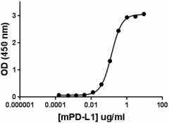
Recombinant mouse PD-1 at 1.0 µg/ml binds to immobilized mouse PD-L1 (Cat. No. 758202) in a dose dependent manner with an ED50 = 0.1 - 0.5 µg/ml.
Programmed death-1 (PD-1) was initially isolated from apoptosis-induced cells by subtractive hybridization. It is a type I transmembrane protein, and its extracellular region consists of a single Ig-like variable (IgV) domain. The amino acid sequence of mouse PD-1 is 59.4 % identical to the human counterpart, and a putative tyrosine kinase-association motif is well conserved. PD-1 is an inhibitory molecule expressed by activated B and T cells and has been implicated in immune tolerance. Mice deficient in PD-1 develop lupus-like proliferative arthritis and glomerulonephritis. PD-1 ligand 1 (PD-L1) and then PD-1 ligand 2 (PD-L2) are the ligands of PD-1. Binding of PD-1 to its ligand PD-L1 leads to the inhibition of T cell receptor-mediated lymphocyte proliferation and cytokine secretion. PD-1/PD-L1 interaction suppresses immune responses against autoantigens and tumors and plays an important role in the maintenance of peripheral immune tolerance. Antibodies against PD-1 or PD-L1 leads to increased antitumor immunity. PD-1 and its ligand PD-L1 are overexpressed in many autoimmune diseases, including inflammatory bowel disease, rheumatoid arthritis, and type 1 diabetes. Interactions between PD-1 and PD-L1 promote tolerance by blocking the TCR-induced stop signal. PD-L1 expression by tumor cells may induce and maintain regulatory T cells in the tumor, allowing tumor progression by enhanced suppression of antitumor T-cell responses. PD-1, PD-L1 and PD-L2 also have soluble forms aside from their membrane bound form.
Product DetailsProduct Details
- Source
- Mouse PD-1, amino acids (Leu25-Gln167, Accession # NP_032824) was expressed in 293E cells. The carboxy terminus contains SRIEGRMD-hIgG-Fc.
- Molecular Mass
- The 382 amino acid recombinant protein has a predicted molecular mass of approximately 43.0 kD. The DTT-reduced and non-reduced protein migrate at approximately 60 kD and 120 kD respectively by SDS-PAGE. The predicted N-terminal amino acid is Leu.
- Purity
- >95% by SDS-PAGE gel as determined by Coomassie stained SDS-PAGE.
- Formulation
- 0.22 µm filtered protein solution is in PBS pH 7.2.
- Endotoxin Level
- Less than 0.01 EU per µg cytokine as determined by the LAL method.
- Concentration
- 10 µg size is bottled at 100 µg/mL
- Storage & Handling
- Unopened vial can be stored between 2°C and 8°C for up to 2 weeks, at -20°C for up to six months, or at -70°C or colder until the expiration date. For maximum results, quick spin vial prior to opening. The protein can be aliquoted and stored at -20°C or colder. Stock solutions can also be prepared at 50 - 100 µg/mL in appropriate sterile buffer, carrier protein such as 0.2 - 1% BSA or HSA can be added when preparing the stock solution. Aliquots can be stored between 2°C and 8°C for up to one week and stored at -20°C or colder for up to 3 months. Avoid repeated freeze/thaw cycles.
- Activity
- Measured by functional ELISA. Immobilized mouse recombinant PD-L1 at different concentrations binds mouse recombinant PD-1 when it is used at 1 µg/ml. ED50 = 0.1 - 0.5 µg/ml.
- Application
-
Bioassay
- Application Notes
-
BioLegend carrier-free recombinant proteins provided in liquid format are shipped on blue-ice. Our comparison testing data indicates that when handled and stored as recommended, the liquid format has equal or better stability and shelf-life compared to commercially available lyophilized proteins after reconstitution. Our liquid proteins are verified in-house to maintain activity after shipping on blue ice and are backed by our 100% satisfaction guarantee. If you have any concerns, contact us at tech@biolegend.com.
- Product Citations
-
Antigen Details
- Structure
- Homodimer
- Distribution
-
Antigen-stimulated T and B cells, regulatory T cells, follicular T and B cells, dendritic cells, and monocytes.
- Function
- PD-1 downregulates immune responses, is involved in immune tolerance. It is induced in activated T cells and B cells.
- Interaction
- APC, monocytes, dendritic cells, stromal cell, cancer cells
- Ligand/Receptor
- B7-H1 (PD-L1, CD274) and B7-DC (PD-L2, CD273)
- Biology Area
- Inhibitory Molecules
- Antigen References
-
1. Ishida Y, et al. 1992 EMBO J. 11:3887.
2. Fife BT, et al. 2009 Nature Immunol. 10(11):1185-92.
3. Freeman GJ, et al. 2000 J. Exp. Med. 192:1027.
4. Latchman Y, et al. 2001 Nat. Immunol. 2001 2:261.
5. Iwai Y, et al. 2002 Proc. Natl. Acad. Sci. USA. 99(19):12293-7.
6. Dai S, et al. 2014 Cell. Immunol. 290:72.
7. Tan S, et al. 2017 Nature Commun. 8:14369. - Gene ID
- 18566 View all products for this Gene ID
- UniProt
- View information about PD-1 on UniProt.org
Related Pages & Pathways
Pages
Related FAQs
- Why choose BioLegend recombinant proteins?
-
• Each lot of product is quality-tested for bioactivity as indicated on the data sheet.
• Greater than 95% Purity or higher, tested on every lot of product.
• 100% Satisfaction Guarantee for quality performance, stability, and consistency.
• Ready-to-use liquid format saves time and reduces challenges associated with reconstitution.
• Bulk and customization available. Contact us.
• Learn more about our Recombinant Proteins. - How does the activity of your recombinant proteins compare to competitors?
-
We quality control each and every lot of recombinant protein. Not only do we check its bioactivity, but we also compare it against other commercially available recombinant proteins. We make sure each recombinant protein’s activity is at least as good as or better than the competition’s. In order to provide you with the best possible product, we ensure that our testing process is rigorous and thorough. If you’re curious and eager to make the switch to BioLegend recombinants, contact your sales representative today!
- What is the specific activity or ED50 of my recombinant protein?
-
The specific activity range of the protein is indicated on the product datasheets. Because the exact activity values on a per unit basis can largely fluctuate depending on a number of factors, including the nature of the assay, cell density, age of cells/passage number, culture media used, and end user technique, the specific activity is best defined as a range and we guarantee the specific activity of all our lots will be within the range indicated on the datasheet. Please note this only applies to recombinants labeled for use in bioassays. ELISA standard recombinant proteins are not recommended for bioassay usage as they are not tested for these applications.
- Have your recombinants been tested for stability?
-
Our testing shows that the recombinant proteins are able to withstand room temperature for a week without losing activity. In addition the recombinant proteins were also found to withstand four cycles of freeze and thaw without losing activity.
- Does specific activity of a recombinant protein vary between lots?
-
Specific activity will vary for each lot and for the type of experiment that is done to validate it, but all passed lots will have activity within the established ED50 range for the product and we guarantee that our products will have lot-to-lot consistency. Please conduct an experiment-specific validation to find the optimal ED50 for your system.
- How do you convert activity as an ED50 in ng/ml to a specific activity in Units/mg?
-
Use formula Specific activity (Units/mg) = 10^6/ ED50 (ng/mL)

 Login / Register


Follow Us