- Regulatory Status
- RUO
- Other Names
- Please refer to individual product datasheets.
- Ave. Rating
- Submit a Review
- Product Citations
- publications

-

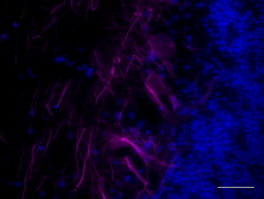
IHC staining of Alexa Fluor® 647 anti-Neurofilament L (NF-L) antibody (clone NFL3) on formalin-fixed paraffin-embedded human brain tissue. Following antigen retrieval using Sodium Citrate H.I.E.R (Cat. No. 928602), the tissue was incubated with 5 µg/ml of the primary antibody overnight at 4°C. The image was captured with a 40X objective. Scale bar: 50 µm -

IHC staining of Alexa Fluor® 488 anti-Neurofilament H & M (NF-H/NF-M), Phosphorylated antibody (clone SMI 310) on formalin-fixed paraffin-embedded mouse midbrain tissue. Following antigen retrieval using Retrieve-All Antigen Unmasking System 3 (Cat. No. 927601), the tissue was incubated with 10 µg/mL of the primary antibody overnight at 4°C. Nuclei were counterstained with DAPI, and the slides were mounted with ProLong™ Gold Antifade Mountant. The image was captured with a 40X objective. Scale Bar: 50 µm -

IHC staining of Alexa Fluor® 488 anti-Neurofilament H (NF-H), Nonphosphorylated antibody (Clone SMI 32) on formalin-fixed paraffin-embedded normal rat brain tissue. Following antigen retrieval using Retrieve-All Antigen Unmasking System 3, the tissue was incubated with the primary antibody at 5 µg/ml overnight at 4°C. Image was captured with a 40X objective. -

IHC staining of Alexa Fluor® 594 anti-Neurofilament H (NF-H), Phosphorylated (clone SMI 31) on formalin-fixed paraffin-embedded rat brain tissue. Following antigen retrieval using Retrieve-All Antigen Unmasking System 3 (Cat. No. 927701), the tissue was incubated with 5 µg/mL of the primary antibody for 1 hour at room temperature. The image was captured with a 40x objective. -

SH-SY5Y neuroblastoma cells were fixed with 4% paraformaldehyde, and then permeabilized with 0.1% Triton x100 for 20 minutes. Cells were blocked with 2% Normal Goat Serum for 30 minutes at room temperature, then incubated with Alexa Fluor® 594 conjugated anti-NF-H/M Hypophosphorylated antibody at 5 µg/mL for 3 hours at room temperature. Nuclei were stained with DAPI (bottom panel, blue). Images were captured with a 40X objective. -

Immunofluorescence staining of anti-Neurofilament H & M (NF-H/NF-M), Hypophosphorylated (SMI 35) conjugated to Alexa Fluor® 594 on formalin-fixed, paraffin-embedded rat brain tissue. Following antigen retrieval using Retrieve-All Antigen Unmasking Solution, the tissue was incubated with the Alexa Fluor® 594-conjugated antibody at 5 µg/mL for one hour at room temperature. The image was captured with a 40X objective.
| Cat # | Size | Price | Quantity Check Availability | Save | ||
|---|---|---|---|---|---|---|
| 899912 | 1 kit | £366 | ||||
The Neurofilament L/M/H Antibody Sampler Kit offers flexibility for sampling and detection of neurofilaments under normal and pathophysiological conditions. This selection of antibodies allows visualization of neuronal axons, dendrites, and cell bodies depending on the clone utilized. Clone SMI 31 reacts primarily with axons while clone SMI 32 visualizes neuronal cell bodies, axons, and dendrites. Clones SMI 35 and SMI 310 generally react with axons. Clone SMI 35 may be used to detect early neuronal cell pathology and intraneuronal neurofibrillary tangles in Alzheimer’s disease (AD). Clone SMI 310 demonstrates strong reaction with extraneuronal (ghost) neurofibrillary tangles in AD. These antibodies provide an easy and rapid solution for multiplexing in ICC or IHC applications.
Product DetailsKit Contents
- Kit Contents
-
Specificity Format Clone Size Reactivity Isotype Anti-Neurofilament L (NF-L) Alexa Fluor® 647 NFL3 25 μg Human, Mouse, Rat Mouse IgG1, κ Anti-Neurofilament H & M (NF-H/NF-M), Phosphorylated Alexa Fluor® 488 SMI 310 25 μg Human, Mouse, Rat Mouse IgG1, κ Anti-Neurofilament H (NF-H), Nonphosphorylated Alexa Fluor® 488 SMI 32 25 μg Rat, mammalian, mouse Mouse IgG1, κ Anti-Neurofilament H (NF-H), Phosphorylated Alexa Fluor® 594 SMI 31 25 μg Human, Mouse, Rat Mouse IgG1, κ Anti-Neurofilament H & M (NF-H/NF-M), Hypophosphorylated Alexa Fluor® 594 SMI 35 25 μg Human, Mouse, Rat Mouse IgG1, κ * For detailed information about each specificity, please refer to the datasheets of the individual products.
Product Details
- Formulation
- Please refer to individual product datasheets.
- Preparation
- Please refer to individual product datasheets.
- Storage & Handling
- Upon receipt, store undiluted at 2-8°C.
- Application
-
IHC-P, ICC - Quality tested
- Recommended Usage
-
Each lot of this antibody is quality control tested by formalin-fixed paraffin-embedded immunohistochemical staining. For immunohistochemistry, the suggested uses of these reagents are as follows:
Anti-Neurofilament L (NF-L): 5.0 - 10 µg/ml
Anti-Neurofilament H & M (NF-H/NF-M), Phosphorylated: 5.0 - 10 µg/ml
Anti-Neurofilament H (NF-H), Nonphosphorylated: 1.0 - 5.0 µg/ml
Anti-Neurofilament H (NF-H), Phosphorylated: 1.0 - 5.0 µg/ml
For immunocytochemistry, the suggested uses of these reagents are as follows:
Anti-Neurofilament H & M (NF-H/NF-M), Hypophosphorylated: 5.0 - 10 µg/ml
It is recommended that the reagent be titrated for optimal performance for each application. - Application Notes
-
For verified or reported applications for these antibodies, please see individual product datasheets.
Antigen Details
- Biology Area
- Cell Biology, Neuroscience, Neuroscience Cell Markers
- Antigen References
-
Please refer to individual product data sheets for antigen references.
- Gene ID
- 4744 View all products for this Gene ID 4747 View all products for this Gene ID

 Login / Register


Follow Us