- Clone
- A20005D (See other available formats)
- Regulatory Status
- RUO
- Other Names
- Linker For Activation Of T Cells; P36-38
- Isotype
- Mouse IgG2b, κ
- Ave. Rating
- Submit a Review
| Cat # | Size | Price | Quantity Check Availability | Save | ||
|---|---|---|---|---|---|---|
| 946601 | 25 µL | £109 | ||||
| 946602 | 100 µL | £254 | ||||
LAT, or Linker for Activation of T cells, is a transmembrane protein expressed mainly in T cells and a limited number of other immune cells such as natural killer cells, mast cells, and immature B cells. As an adaptor protein, phosphorylated LAT facilitates the recruitment of other signaling proteins and functions as a nucleating site for multiprotein signaling complexes. These signaling complexes can propagate TCR, or T Cell antigen Receptors, and activate downstream effectors, which trigger T cell proliferation or cytokine expression.
LAT (Tyr171) is one of the nine tyrosines in LAT conserved between human, mice, and rat. LAT (Tyr171) is necessary for the activation of PI 3-kinase, a protein that regulates the recruitment of other proteins that facilitate in T-cell receptor signal transduction such as PLC-gamma1 and Sos1. When LAT (Tyr171) and (Tyr191) is phosphorylated by ZAP-70, Gads can bind directly to LAT (Tyr171) and (Tyr191) and mediate the recruitment of SLP-76.
Product Details
- Verified Reactivity
- Human, Mouse
- Antibody Type
- Monoclonal
- Host Species
- Mouse
- Immunogen
- Synthetic peptide corresponding to human LAT phosphorylated at Tyr 171
- Formulation
- Phosphate Buffer, 0.5% BSA, 0.09% NaN3
- Preparation
- The antibody was purified by affinity chromatography.
- Concentration
- 0.05 mg/mL
- Storage & Handling
- The antibody solution should be stored undiluted between 2°C and 8°C.
- Application
-
WB - Quality tested
ICC, ICFC - Verified - Recommended Usage
-
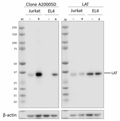
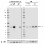
Each lot of this antibody is quality control tested by western blotting. For western blotting, the suggested dilution of this reagent is 1000 - 1:5000. For immunocytochemistry, a dilution of 1:10 is recommended. For intracellular flow cytometric staining, the suggested dilution of this reagent is 1:20 per million cells in 100 µL volume. It is recommended that the reagent be titrated for optimal performance for each application.
- Application Notes
-
When this clone is used in WB at a dilution lower than 1:1000 it may show non-specific bands at 50 kD and above.
This clone has been tested by ICC on H2O2 treated and untreated Jurkat, with three fix and permeabilization methods (100% methanol, 4% PFA plus methanol and 4% PFA plus Triton X-100). All three methods can be used for ICC staining, but the PFA plus Triton X-100 fixation/permeabilization method produced the best staining.
This clone has only been tested on True-PhosTM perm buffer (Cat. No. 425401) by ICFC - RRID
-
AB_2904456 (BioLegend Cat. No. 946601)
AB_2904456 (BioLegend Cat. No. 946602)
Antigen Details
- Structure
- LAT is a 262 amino acid protein with a predicted molecular weight of 36-38 kD.
- Distribution
-
Plasma membrane/T Cells
- Function
- Plasma membrane/T Cells
- Cell Type
- Mast cells, NK cells, T cells
- Biology Area
- Cell Biology, Immunology, Signal Transduction
- Molecular Family
- Phospho-Proteins
- Antigen References
-
- Balagopalan L. et al., 2010, Cold Spring Harb Perspect Biol., 2(8): a005512
- Balagopalan L. et al., 2020, PLoS One, 15(2): e0229036
- Lin J. and Weiss A., 2001, J Biol Chem., 276(31): 29588-95
- Paz et. al., 2001, Biochem J., 356, 461-71
- Zhang W. et al., 2000, J Biol Chem., 275(30): 23355-61
- Gene ID
- 27040 View all products for this Gene ID
- UniProt
- View information about LAT Phospho Tyr171 on UniProt.org
Related FAQs
Other Formats
View All LAT Phospho (Tyr171) Reagents Request Custom Conjugation| Description | Clone | Applications |
|---|---|---|
| Purified anti-LAT Phospho (Tyr171) | A20005D | WB,ICC,ICFC |
| Alexa Fluor® 647 anti-LAT Phospho (Tyr171) | A20005D | ICFC |
| PE anti-LAT Phospho (Tyr171) | A20005D | ICFC |
Compare Data Across All Formats
This data display is provided for general comparisons between formats.
Your actual data may vary due to variations in samples, target cells, instruments and their settings, staining conditions, and other factors.
If you need assistance with selecting the best format contact our expert technical support team.
-
Purified anti-LAT Phospho (Tyr171)
Whole cell extracts (15 μg protein) from serum-starved Jurka... 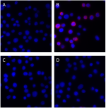
Serum-starved Jurkat cells untreated (lower) and treated (up... 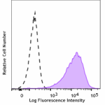
Jurkat cells treated (filled histogram, positive control) wi... -
Alexa Fluor® 647 anti-LAT Phospho (Tyr171)
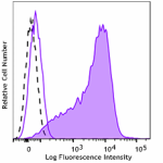
Jurkat cells untreated (negative control, open histogram) or... 
Peripherical blood mononuclear cells (PBMC) untreated (left)... -
PE anti-LAT Phospho (Tyr171)
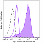
Jurkat cells untreated (negative control, open histogram), o... 
Peripherical blood mononuclear cells (PBMC) untreated (left)...

 Login / Register


Follow Us