 Login / Register


- Clone
- MCA-5C10 (See other available formats)
- Regulatory Status
- RUO
- Other Names
- GFAP, Glial fibrillary acidic protein
- Isotype
- Mouse IgG1, κ
- Ave. Rating
- Submit a Review
- Product Citations
- publications

-

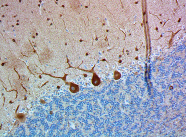
IHC staining of purified anti-GFAP antibody (clone MCA-5C10) on formalin-fixed paraffin-embedded human brain tissue. Following antigen retrieval using Sodium Citrate H.I.E.R., the tissue was incubated with 2 µg/ml of the primary antibody for 60 minutes at room temperature. BioLegend's Ultra-Streptavidin (USA) HRP kit (Multi-Species, DAB, Cat. No. 929901) was used for detection followed by hematoxylin counterstaining, according to the protocol provided. The image was captured with a 40X objective. Scale bar: 50 µm -

IHC staining of purified anti-GFAP antibody (clone MCA-5C10) on formalin-fixed paraffin-embedded mouse brain tissue. Following antigen retrieval using Sodium Citrate H.I.E.R., the tissue was incubated with 2 µg/ml of the primary antibody for 60 minutes at room temperature. BioLegend's Ultra-Streptavidin (USA) HRP kit (Multi-Species, DAB, Cat. No. 929901) was used for detection followed by hematoxylin counterstaining, according to the protocol provided. The image was captured with a 40X objective. Scale bar: 50 µm -

IHC staining of purified anti-GFAP antibody (clone MCA-5C10) on formalin-fixed paraffin-embedded rat brain tissue. Following antigen retrieval using Sodium Citrate H.I.E.R., the tissue was incubated with 2 µg/ml of the primary antibody for 60 minutes at room temperature. BioLegend's Ultra-Streptavidin (USA) HRP kit (Multi-Species, DAB, Cat. No. 929901) was used for detection followed by hematoxylin counterstaining, according to the protocol provided. The image was captured with a 40X objective. Scale bar: 50 µm -

IHC staining of purified anti-GFAP antibody (clone MCA-5C10) on formalin-fixed paraffin-embedded rat brain tissue. Following antigen retrieval using Sodium Citrate H.I.E.R. (Cat. No.928502), the tissue was incubated with 2 µg/mL of the primary antibody overnight at 4°C, followed by incubation with Alexa Fluor® 594 goat anti-mouse IgG (Cat. No. 405326) for one hour at room temperature. The slide was mounted with Fluoromount-G with DAPI. The image was captured with a 40X objective. Scale Bar: 50 µm -

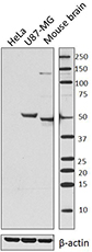
Western blot of purified anti-GFAP antibody (clone MCA-5C10). Lane 1: Molecular weight marker; Lane 2: 20 µg of human brain lysate; Lane 3: 20 µg of mouse brain membrane lysate; Lane 4: 40 µg of mouse brain lysate; Lane 5: 20 µg rat brain membrane lysate. The blot was incubated with 0.2 µg/mL of the primary antibody overnight at 4°C, followed by incubation with HRP labeled goat anti-mouse IgG (Cat. No. 405306). Enhanced chemiluminescence was used as the detection system.
| Cat # | Size | Price | Quantity Check Availability | Save | ||
|---|---|---|---|---|---|---|
| 801103 | 25 µg | 85€ | ||||
Glial Fibrillary Acidic Protein (GFAP) is a major fibrous protein of multiple sclerosis plaques. It was subsequently found to be a member of the 10nm or intermediate filament protein family, specifically the intermediate filament protein family Class III, which also includes peripherin, desmin and vimentin. GFAP is strongly and specifically expressed in astrocytes and certain other astroglia in the central nervous system, in satellite cells in peripheral ganglia, and in non-myelinating Schwann cells in peripheral nerves. Astrocytes respond to many damage and disease states resulting in "astrogliosis" or the presence of a "glial response". GFAP antibodies are widely used to see the reactive astrocytes which form part of this response, since reactive astrocytes stain much more strongly with GFAP antibodies than normal astrocytes. GFAP also forms a major component of the so-called glial scar, an astrocyte rich structure apparently forming part of the barrier to nerve fiber regeneration following damage in the central nervous system. Neural stem cells frequently strongly express GFAP. Antibodies to GFAP are therefore very useful as markers of normal and reactive astrocytic cells and neural stem cells. This antibody was raised against a preparation of purified pig spinal core GFAP.
Product DetailsProduct Details
- Verified Reactivity
- Human, Mouse, Rat
- Antibody Type
- Monoclonal
- Host Species
- Mouse
- Formulation
- PBS, 50% glycerol, 5mM NaN3 as supplied by vendor.
- Preparation
- The antibody was purified by affinity chromatography.
- Concentration
- 0.5 mg/ml
- Storage & Handling
- Store at 4°C for up to 3 months. For longer term storage aliquot into small volumes and store at -20°C for up to one year.
- Application
-
IHC-P - Quality tested
WB - Verified
ICC, IHC-F - Reported in the literature, not verified in house - Recommended Usage
-
Each lot of this antibody is quality control tested by formalin-fixed paraffin-embedded immunohistochemical staining. For immunohistochemistry, a concentration range of 2.0 - 5.0 µg/mL for chromogenic staining and 2.0 - 10 µg/mL for fluorescent staining is suggested. For Western blotting, the suggested use of this reagent is 0.1 - 0.2 µg per mL. It is recommended that the reagent be titrated for optimal performance for each application.
- Application References
-
- Filipowicz AR, et al. 2016. Sci Rep. 6: 32900. PubMed (IHC-P)
- Edamakanti CR, et al. 2018. J. Clin Invest. 128(6):2252 (ICC)
- de Kloet AD, et al. 2016. Brain Struct Funct. 221(2):891 (IHC-F)
- Hawkins KE, et al. 2014. J Neurochem. 129(1):130 (WB)
- Zhao WN, et al. 2012. J Biomol Screen. 17(9):1252 (ICC)
- Product Citations
-
- RRID
-
AB_2810699 (BioLegend Cat. No. 801103)
Antigen Details
- Structure
- GFAP is a 432 amino acid protein with a molecular mass of ~50 kD
- Distribution
-
Tissue distribution: GFAP is expressed by numerous cell types of the central nervous system (CNS) including astrocytes, ependymal cells, and Bergmann glia cells (protoplasmic astrocyte). GFAP is expressed in cells lacking fibronectin.
Cellular distribution: Cytoskeleton and cytosol - Function
- GFAP is a class-III intermediate filament and a structural constituent of the cytoskeleton. It is a cell-specific marker that is used to distinguish astrocytes from other glial cells during the development of the CNS
- Antigen References
-
- van Bodegraven EJ, et al. 2019. Glia. 10.1002/glia.23594.
- Pekny M, et al. 2019. Neurosci Lett. 689:45.
- Hol EM, et al. 2017. Cold Spring Harb Perspect Biol. 9(12).
- Gene ID
- 2670 View all products for this Gene ID 14580 View all products for this Gene ID 24387 View all products for this Gene ID
- UniProt
- View information about GFAP on UniProt.org

Other Formats
View All GFAP Reagents Request Custom Conjugation| Description | Clone | Applications |
|---|---|---|
| Purified anti-GFAP | MCA-5C10 | IHC-P,WB,ICC,IHC-F |
Customers Also Purchased
Compare Data Across All Formats
This data display is provided for general comparisons between formats.
Your actual data may vary due to variations in samples, target cells, instruments and their settings, staining conditions, and other factors.
If you need assistance with selecting the best format contact our expert technical support team.
-
Purified anti-GFAP

IHC staining of purified anti-GFAP antibody (clone MCA-5C10)... 
IHC staining of purified anti-GFAP antibody (clone MCA-5C10)... 
IHC staining of purified anti-GFAP antibody (clone MCA-5C10)... 
IHC staining of purified anti-GFAP antibody (clone MCA-5C10)... 
Western blot of purified anti-GFAP antibody (clone MCA-5C10)...


Follow Us