- Clone
- ME-9F1 (See other available formats)
- Regulatory Status
- RUO
- Other Names
- S-Endo 1 antigen, MUC18, MCAM, Mel-CAM, A32 antigen
- Isotype
- Rat IgG2a, κ
- Ave. Rating
- Submit a Review
- Product Citations
- publications
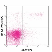
-

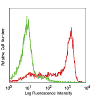
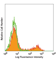
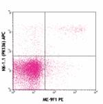
C57BL/6 splenocytes stained with ME-9F1 PE and NK-1.1 (PK136) APC -

C57BL/6 splenocytes stained with rat IgG2a (RTK2758) PE isotype control and NK-1.1 (PK136) APC -

C57BL/6 mouse frozen spleen section was fixed with 4% paraformaldehyde (PFA) for 10 minutes at room temperature and blocked with 5% FBS for 30 minutes at room temperature. Then the section was stained with 10 µg/ml of CD146 (clone ME-9F1) purified, and CD3ε (clone 500A2) Alexa Fluor® 594 (green) overnight at 4°C, followed by 2.5 µg/ml of anti-rat IgG (clone Poly4054) Alexa Fluor® 647 (red) for 2 hours at room temperature. Nuclei were counterstained with DAPI (blue). The image was captured by 10X objective.
| Cat # | Size | Price | Quantity Check Availability | Save | ||
|---|---|---|---|---|---|---|
| 134701 | 50 µg | 81€ | ||||
| 134702 | 200 µg | 249€ | ||||
CD146, also known as melanoma cell adhesion molecule (MCAM or Mel-CAM), MUC18, S-Endo1, and A32 antigen, is an integral membrane glycoprotein that belongs to the Ig superfamily. CD146 is strongly expressed by murine vascular endothelial cells. It is expressed on about 30% of neutrophils and 60% of NK cells. Unlike in humans, CD146 is undetectable on monocytes, dendritic cells, T cells, NKT cells, B cells, or smooth muscle cells in mouse. It has been reported that an increase in CD146 expression is associated with NK cell maturation. Combined with using CD27 and CD11b staining, CD146 may be an alternative marker to detect final stages of NK cell maturation and define NK cell subsets. CD146+ NK cells were found to be less cytotoxic and to produce less IFNγ than CD146- NK cells upon stimulation with target cells or activating antibodies. The role of CD146 on NK cell migration has yet to be investigated. The identification of CD146 ligand(s) will be crucial to address this issue.
Product DetailsProduct Details
- Verified Reactivity
- Mouse
- Antibody Type
- Monoclonal
- Host Species
- Rat
- Immunogen
- Endothelial cell line TME-3H3
- Formulation
- Phosphate-buffered solution, pH 7.2, containing 0.09% sodium azide.
- Preparation
- The antibody was purified by affinity chromatography.
- Concentration
- 0.5 mg/ml
- Storage & Handling
- The antibody solution should be stored undiluted between 2°C and 8°C.
- Application
-
FC - Quality tested
IHC-F - Verified - Recommended Usage
-
Each lot of this antibody is quality control tested by immunofluorescent staining with flow cytometric analysis. For flow cytometric staining, the suggested use of this reagent is ≤ 0.25 µg per million cells in 100 µl volume. For immunohistochemistry, a concentration range of 5.0 - 10.0 µg/ml is suggested. It is recommended that the reagent be titrated for optimal performance for each application.
- Application References
-
- Schrage A, et al. 2008. Histochem. Cell. Biol. 129:441.
- Birbrair A, et al. 2013. Am J Physiol Cell Physiol. 305:1098. PubMed
- Product Citations
-
- RRID
-
AB_1732002 (BioLegend Cat. No. 134701)
AB_1731991 (BioLegend Cat. No. 134702)
Antigen Details
- Structure
- CD146 is a transmembrane glycoprotein and belongs to the Ig superfamily of cell adhesion molecules.
- Distribution
-
CD146 is strongly expressed by murine vascular endothelial cells, a subset of NK1.1+ cells, and neutrophils.
- Function
- CD146 has been reported to be functionally relevant for endothelial cell adhesion, angiogenesis, and NK cell function.
- Ligand/Receptor
- Unidentified ligand.
- Cell Type
- Endothelial cells, Mesenchymal Stem Cells, Neutrophils
- Biology Area
- Cell Biology, Immunology, Neuroscience, Neuroscience Cell Markers, Stem Cells
- Molecular Family
- Adhesion Molecules, CD Molecules
- Antigen References
-
1. Despoix N, et al. 2008. Eur. J. Immunol. 38:2855.
2. Sorrentino A, et al. 2008. Exp. Hematol. 36:1035.
3. Bardin N, et al. 2009. Arterioscler. Thromb. Vasc. Biol. 29:746. - Gene ID
- 84004 View all products for this Gene ID
- UniProt
- View information about CD146 on UniProt.org
Related FAQs
Other Formats
View All CD146 Reagents Request Custom Conjugation| Description | Clone | Applications |
|---|---|---|
| PerCP/Cyanine5.5 anti-mouse CD146 | ME-9F1 | FC |
| Purified anti-mouse CD146 | ME-9F1 | FC,IHC-F |
| PE anti-mouse CD146 | ME-9F1 | FC |
| FITC anti-mouse CD146 | ME-9F1 | FC |
| Alexa Fluor® 488 anti-mouse CD146 | ME-9F1 | FC,IHC-F |
| APC anti-mouse CD146 | ME-9F1 | FC |
| PE/Cyanine7 anti-mouse CD146 | ME-9F1 | FC |
| Alexa Fluor® 647 anti-mouse CD146 | ME-9F1 | FC,IHC-F,3D IHC |
| Biotin anti-mouse CD146 | ME-9F1 | FC |
| Alexa Fluor® 594 anti-mouse CD146 | ME-9F1 | IHC-F,3D IHC,FC |
Customers Also Purchased
Compare Data Across All Formats
This data display is provided for general comparisons between formats.
Your actual data may vary due to variations in samples, target cells, instruments and their settings, staining conditions, and other factors.
If you need assistance with selecting the best format contact our expert technical support team.
-
PerCP/Cyanine5.5 anti-mouse CD146
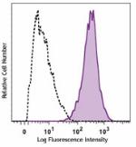
bEND.3 cells (mouse endothelial cells) were stained with CD1... -
Purified anti-mouse CD146
C57BL/6 splenocytes stained with ME-9F1 PE and NK-1.1 (PK136... 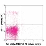
C57BL/6 splenocytes stained with rat IgG2a (RTK2758) PE isot... 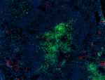
C57BL/6 mouse frozen spleen section was fixed with 4% parafo... -
PE anti-mouse CD146

C57BL/6 splenocytes stained with ME-9F1 PE and NK-1.1 (PK136... 
C57BL/6 splenocytes stained with rat IgG2a (RTK2758) PE isot... -
FITC anti-mouse CD146
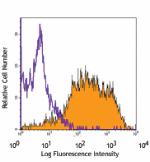
bEND.3 cells (mouse endothelial cells) stained with ME-9F1 F... -
Alexa Fluor® 488 anti-mouse CD146
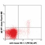
C57BL/6 splenocytes stained with anti-mouse NK-1.1 (PK136) A... 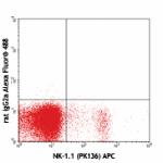
C57BL/6 splenocytes stained with NK-1.1 (PK136) APC and rat ... 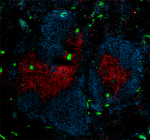
C57BL/6 mouse frozen spleen section was fixed with 4% parafo... -
APC anti-mouse CD146
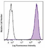
Mouse endothelial cells were stained with CD146 (clone ME-9F... -
PE/Cyanine7 anti-mouse CD146
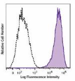
bEnd.3, mouse endothelial cells were stained with CD146 (clo... -
Alexa Fluor® 647 anti-mouse CD146
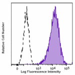
bEnd.3 cells (mouse endothelial cells) were stained with CD1... 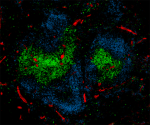
C57BL/6 mouse frozen spleen section was fixed with 4% parafo... 
Paraformaldehyde-fixed (4%), 500 μm-thick mouse spleen secti... -
Biotin anti-mouse CD146
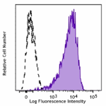
bEND.3 cells (mouse endothelial cells) were stained with bio... -
Alexa Fluor® 594 anti-mouse CD146

C57BL/6 mouse frozen spleen section was fixed with 4% parafo... 
Paraformaldehyde-fixed (4%), 500 μm-thick mouse spleen secti... 
C57BL/6 mouse splenocytes were stained with anti-mouse NK-1....
 Login / Register




Follow Us