- Clone
- 10C3 (See other available formats)
- Regulatory Status
- RUO
- Other Names
- Macrophage migration inhibitory factor (MMIF), Glycosylation inhibiting factor (GIF), GLIF
- Isotype
- Mouse IgG2b, κ
- Ave. Rating
- Submit a Review
- Product Citations
- publications

-

-

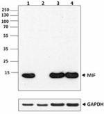
Western blot analysis of THP-1 (lane 1), Raw264.7(lane 2), PC-3(lane 3) and Jurkat(lane 4) cells using anti-MIF antibody (10C3). GAPDH antibody (poly6314) was used as loading control.
| Cat # | Size | Price | Quantity Check Availability | Save | ||
|---|---|---|---|---|---|---|
| 525501 | 50 µg | $124 | ||||
| 525502 | 500 µg | $358 | ||||
Human macrophage migration inhibitory factor (MIF) is a 12.5 kD, 115 amino acid, non-glycosylated polypeptide expressed by multiple cell types, including activated T cells, macrophages, eosinophils, epithelial cells, and endothelial cells. MIF plays many roles in biological processes such as catalytic activity, immunity, endocrine regulation, signal modulation, and inflammation.
MIF is expressed in malignant cells including lung, liver, breast, colon, and prostate tumors. Studies suggest that MIF might serve as a molecular link between chronic inflammation and cancer.
Recombinant MIF consists of a mixture of monomers, dimers, and trimers. The physiologically active forms are believed to be predominantly dimeric and trimeric forms.
Product Details
- Verified Reactivity
- Human
- Antibody Type
- Monoclonal
- Host Species
- Mouse
- Immunogen
- Recombinant human MIF with a His tag
- Formulation
- Phosphate-buffered solution, pH 7.2, containing 0.09% sodium azide.
- Preparation
- The antibody was purified by affinity chromatography.
- Concentration
- 0.5 mg/ml
- Storage & Handling
- The antibody solution should be stored undiluted between 2°C and 8°C.
- Application
-
ELISA Capture - Quality tested
WB - Verified - Recommended Usage
-
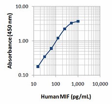
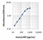
Each lot of this protein is quality control tested by ELISA assay. A concentration of 4 µg/ml of the capture antibody was utilized to generate the example standard curve. It is recommended that each lot of reagent be titrated for optimal performance for each application.
- Application Notes
-
ELISA: To measure human MIF, the purified 10C3 antibody (Cat. No. 525501) is useful as the capture antibody in a sandwich ELISA assay, when used in conjunction with the biotinylated 10C3 antibody (Cat. No. 525503) as the detection antibody.
Additional reported applications (for the relevant formats) include: functional assay1 and neutralization1. The LEAF™ purified antibody (Endotoxin <0.1 EU/µg, Azide-Free, 0.2 µm filtered) is recommended for functional assays (contact our custom solutions team).
Note: For testing human MIF in serum, plasma or cell culture supernatant, BioLegend's LEGEND MAX™ ELISA Kits (Cat. No. 438407 & 438408) are specially developed and recommended. -
Application References
(PubMed link indicates BioLegend citation) -
- Zhou H, et al. 2009. PLoS ONE. 4:e6087. (FA, Neut)
- Product Citations
-
- RRID
-
AB_2563133 (BioLegend Cat. No. 525501)
AB_2563133 (BioLegend Cat. No. 525502)
Antigen Details
- Structure
- Predominantly a homotrimer
- Distribution
-
Activated T-cells, macrophages, eosinophils, epithelial cells, and endothelial cells
- Function
- Pro-inflammatory cytokine, regulates macrophage function, involved in cancer development, tautomerase activity
- Interaction
- Monocytes and macrophages, mesenchymal cells, B-cells
- Ligand/Receptor
- CD74, CXCR4, CXCR2, CXCR7
- Cell Type
- Endothelial cells, Eosinophils, Epithelial cells, Macrophages, T cells
- Biology Area
- Angiogenesis, Apoptosis/Tumor Suppressors/Cell Death, Cell Biology, Cell Cycle/DNA Replication, Immunology, Innate Immunity, Signal Transduction
- Molecular Family
- Cytokines/Chemokines
- Antigen References
-
1. Zhou H, et al. 2009. PLoS ONE. 4:e6087.
2. Potolicchio I, et al. 2003. J. Biol. Chem. 278:30889.
3. Meyer-Siegler K & Iczkowski K. 2005. BMC Cancer. 5:73.
4. Babu SN, et al. 2012. Asian Pac. J. Cancer Prev. 13:1737. - Gene ID
- 4282 View all products for this Gene ID
- Specificity (DOES NOT SHOW ON TDS):
- MIF
- Specificity Alt (DOES NOT SHOW ON TDS):
- MIF
- App Abbreviation (DOES NOT SHOW ON TDS):
- ELISA Capture,WB
- UniProt
- View information about MIF on UniProt.org
Related Pages & Pathways
Pages
Related FAQs
Other Formats
View All MIF Reagents Request Custom Conjugation| Description | Clone | Applications |
|---|---|---|
| Purified anti-human MIF | 10C3 | ELISA Capture,WB |
| Biotin anti-human MIF | 10C3 | ELISA Detection |
Customers Also Purchased
Compare Data Across All Formats
This data display is provided for general comparisons between formats.
Your actual data may vary due to variations in samples, target cells, instruments and their settings, staining conditions, and other factors.
If you need assistance with selecting the best format contact our expert technical support team.
-
Purified anti-human MIF

Western blot analysis of THP-1 (lane 1), Raw264.7(lane 2), P... 
-
Biotin anti-human MIF

 Login/Register


Follow Us