Add a new dimension to your research by pairing your fluorescent antibodies with our Ce3D™ Tissue Clearing Solution. Designed by academic researchers, this buffer provides unparalleled clarity and insight as you interrogate tissues to produce mesmerizing 3D images.


Tissue clearing reagents, like Ce3D, reduce light scattering by normalizing the refractive index throughout the tissue and make the tissue transparent. Optimal clearing empowers 3D imaging, allowing for a better understanding of spatial composition, phenotypic and subtype identity, and cellular networks.


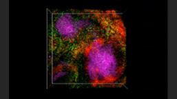
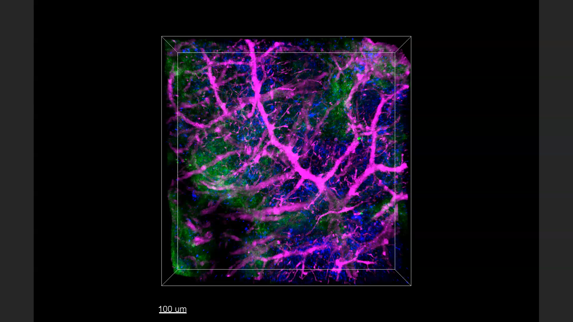
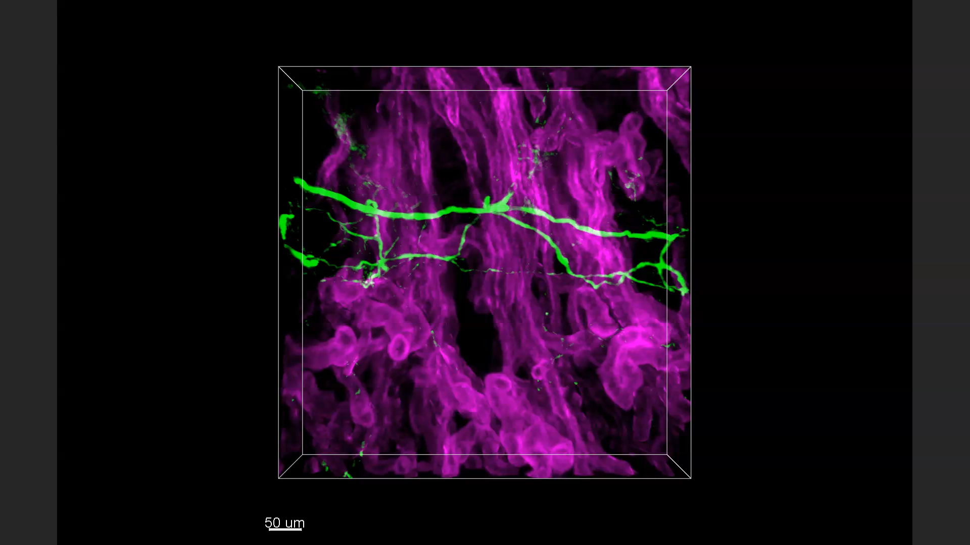
The images above are representative 3D IHC images of 500 μm-thick mouse samples. The sections were optically cleared after immunostaining, mounted in sample chambers, and imaged. From left to right: Mouse spleen: CD68 (green), CD172a (red), CD8a (magenta). Mouse spleen: F4/80 (green), I-A/I-E (red), CD146 (magenta). Mouse kidney: TUBB3 (green), CD326 (magenta).


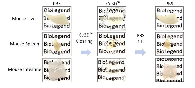
Representative 500 µm-thick mouse tissue sections before and after tissue clearing. Mice were perfused and organs were fixed 4% PFA. Clearing was reversed by immersing tissues in PBS for one hour. 1% PFA is recommended for the best clearing effect. 4% PFA is recommended for optimal epitope preservation and immunostaining.

ProductsHere

Login/Register
Remember me
Forgot your password? Reset Password
Request an Account