- Clone
- 17A12 (See other available formats)
- Regulatory Status
- RUO
- Other Names
- IL-21 Receptor, novel interleukin receptor (NILR), MGC10967
- Isotype
- Mouse IgG1, κ
- Ave. Rating
- Submit a Review
- Product Citations
- publications
-

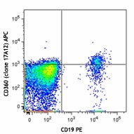
Human peripheral blood lymphocytes were stained with CD19 PE and CD360 (IL-21R) APC (clone 17A12) (top) or mouse IgG1, κ APC isotype control (bottom). -

| Cat # | Size | Price | Quantity Check Availability | Save | ||
|---|---|---|---|---|---|---|
| 359507 | 25 tests | $154 | ||||
| 359508 | 100 tests | $332 | ||||
Human interleukin 21 receptor (IL-21R) is a single pass type I membrane protein and a member of the type I cytokine receptor family. Of the type I cytokine receptors, IL-21R exhibits greatest extracellular homology to the IL-2R beta subunit (i.e., it contains one copy of the WSXWS-containing cytokine-binding domain). Intracellular domains of IL-21R include the Box 1 and Box 2 elements, which are similar to the IL-9R intracellular region. Upon binding IL-21, IL-21R forms a heterodimer with the common gamma subunit (CD132) and induces Jak/Stat signaling. IL-21R is expressed on B cells and at various levels on NK and T cells. IL-21 is a potent immunomodulatory cytokine mainly produced by NKT and CD4 T-cells (particularly the inflammatory Th17 subset) and has pleiotropic effects on both innate and adaptive immune responses. These actions include positive effects such as enhanced proliferation of natural killer (NK) cells and cytotoxic T cells that can destroy virally infected or cancerous cells and direct inhibitory effects on the antigen-presenting function of dendritic cells, and can be proapoptotic for B cells and NK cells. Studies have shown that IL-21 is also an autocrine cytokine that potently induces Th17 differentiation and suppresses Foxp3 expression, and serves as a target for treating inflammatory diseases.
Product DetailsProduct Details
- Verified Reactivity
- Human
- Antibody Type
- Monoclonal
- Host Species
- Mouse
- Immunogen
- Human IL-21R expressing mouse M1-T22 cell line.
- Formulation
- Phosphate-buffered solution, pH 7.2, containing 0.09% sodium azide and BSA (origin USA)
- Preparation
- The antibody was purified by affinity chromatography and conjugated with APC under optimal conditions.
- Concentration
- Lot-specific (to obtain lot-specific concentration and expiration, please enter the lot number in our Certificate of Analysis online tool.)
- Storage & Handling
- The antibody solution should be stored undiluted between 2°C and 8°C, and protected from prolonged exposure to light. Do not freeze.
- Application
-
FC - Quality tested
- Recommended Usage
-
Each lot of this antibody is quality control tested by immunofluorescent staining with flow cytometric analysis. For flow cytometric staining, the suggested use of this reagent is 5 µl per million cells in 100 µl staining volume or 5 µl per 100 µl of whole blood.
- Excitation Laser
-
Red Laser (633 nm)
- Application Notes
-
Additional reported applications (for the relevant formats) include: immunofluorescent surface staining1.
-
Application References
(PubMed link indicates BioLegend citation) -
- Bannantine J, et al. 2011. Front Microbiol. 2:163. (IF)
- RRID
-
AB_2563187 (BioLegend Cat. No. 359507)
AB_2563189 (BioLegend Cat. No. 359508)
Antigen Details
- Structure
- Single pass type I membrane protein, extracellular WSXWS-containing cytokine-binding domain, intracellular Box1 and Box2 domains
- Distribution
-
B cells, various levels on NK cells, T cells, NKT cells, B cells, DCs, macrophages, non-hematopoietic cells, including keratinocytes and fibroblasts
- Ligand/Receptor
- IL-21
- Cell Type
- B cells, Dendritic cells, Fibroblasts, Macrophages, NK cells, NKT cells, T cells, Tregs
- Biology Area
- Immunology, Innate Immunity
- Molecular Family
- CD Molecules, Cytokine/Chemokine Receptors
- Antigen References
-
1. Parrish-Novak J, et al. 2002. J. Leukocyte Biol. 72:856.
2. de Totero D, et al. 2006. Blood 107:3708.
3. Asao H, et al. 2001. J. Immunol. 167:1.
4. Davis ID, et al. 2007. Clin. Cancer Res. 13:6926.
5. Nurieva R. 2007. Nature. 448:480.
6. Dumoutier L, et al. 2000. Proc. Natl. Acad. Sci. USA 97:10144.
7. Ozaki, K, et al. 2000. Proc. Natl. Acad. Sci. USA 97:11439.
8. Parrish-Novak J, et al. 2000. Nature 408:57.
9. Spolski R and Leonard WJ. et al. 2008. Annu. Rev. Immunol. 26:57. - Gene ID
- 50615 View all products for this Gene ID
- UniProt
- View information about CD360 on UniProt.org
Related FAQs
Other Formats
View All CD360 Reagents Request Custom Conjugation| Description | Clone | Applications |
|---|---|---|
| PE anti-human CD360 (IL-21R) | 17A12 | FC |
| APC anti-human CD360 (IL-21R) | 17A12 | FC |
| Brilliant Violet 421™ anti-human CD360 (IL-21R) | 17A12 | FC |
| Brilliant Violet 711™ anti-human CD360 (IL-21R) | 17A12 | FC |
| PE/Cyanine7 anti-human CD360 (IL-21R) | 17A12 | FC |
| TotalSeq™-A0985 anti-human CD360 (IL-21R) Antibody | 17A12 | PG |
| TotalSeq™-C0985 anti-human CD360 (IL-21R) Antibody | 17A12 | PG |
| TotalSeq™-D0985 anti-human CD360 (IL-21R) | 17A12 | PG |
Customers Also Purchased
Compare Data Across All Formats
This data display is provided for general comparisons between formats.
Your actual data may vary due to variations in samples, target cells, instruments and their settings, staining conditions, and other factors.
If you need assistance with selecting the best format contact our expert technical support team.
-
PE anti-human CD360 (IL-21R)
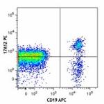
Human peripheral blood lymphocytes were stained with CD19 AP... 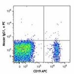
-
APC anti-human CD360 (IL-21R)
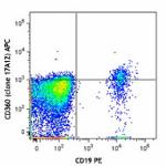
Human peripheral blood lymphocytes were stained with CD19 PE... 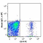
-
Brilliant Violet 421™ anti-human CD360 (IL-21R)
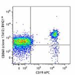
Human peripheral blood lymphocytes were stained with CD19 AP... 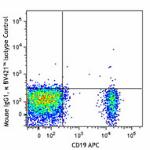
-
Brilliant Violet 711™ anti-human CD360 (IL-21R)
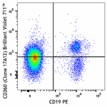
Human peripheral blood lymphocytes were stained with CD19 PE... 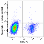
-
PE/Cyanine7 anti-human CD360 (IL-21R)

Human peripheral blood lymphocytes were stained with CD19 AP... -
TotalSeq™-A0985 anti-human CD360 (IL-21R) Antibody
-
TotalSeq™-C0985 anti-human CD360 (IL-21R) Antibody
-
TotalSeq™-D0985 anti-human CD360 (IL-21R)
 Login/Register




Follow Us