- Regulatory Status
- RUO
- Other Names
- TMR Ceramide Golgi Probe
- Ave. Rating
- Submit a Review
- Product Citations
- publications

-

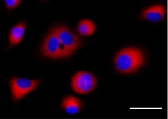
Overlay of live-cell fluorescence imaging of HeLa cells stained with Golgi Detection Probe Red (red). Nuclei were stained with Hoechst 33342 (blue). Images were captured using a 40X objective. Scale bar: 20 µm
| Cat # | Size | Price | Quantity Check Availability | Save | ||
|---|---|---|---|---|---|---|
| 421908 | ea | $500 | ||||
The Golgi apparatus is a complex of vesicles and folded membranes within the cytoplasm of eukaryotic cells. The Golgi apparatus is involved in protein and lipid secretion and intracellular transport. It modifies proteins and lipids that have been built in the endoplasmic reticulum (ER) and prepares them for export outside of the cell. It also plays a significant role in the formation of lysosomes. Golgi Detection Probe Red can selectively bind to the Golgi apparatus and provides a simple and rapid way to perform live-cell fluorescent imaging.
Product DetailsProduct Details
- Verified Reactivity
- Human
- Reported Reactivity
- Mouse, Rat
- Formulation
- 1 vial of Golgi Detection Probe, 1 vial of DMSO
- Preparation
- Please refer to detailed protocol in application notes.
- Storage & Handling
- -20°C
- Application
-
Live cell imaging - Quality tested
- Recommended Usage
-
Please refer to detailed protocol in application notes.
- Application Notes
-
The Golgi Detection Probe Red is a fluorescently labeled ceramide, which selectively binds to the Golgi apparatus and provides a simple method to examine Golgi morphology in live cells. Using the recommended volume of 100 μL per well in a 96-well plate, this product supplies enough probe for 100 tests.
Component:
Lyophilized Golgi Detection Probe, 1 vial
DMSO, 1 vial
Required Materials Not Included:
Phenol-free media or HBSS
Imaging Guidelines:
Ex/Em = 544/570 nm
Fluorescence microscope filter set: Cyanine3/TRITC filter
Flow Guidelines:
Analysis in Cyanine3 or similar channel
Assay Protocol:
1. Prepare the 100X Golgi Detection Probe Stock Solution by adding 100 µL of DMSO to the lyophilized probe. Gently mix until dissolved.
Note: Store the unused Golgi Detection Probe Stock Solution at -20°C in single use aliquots to avoid freeze thaw cycles.
2. Allow the 100X Golgi Detection Probe Stock Solution to reach room temperature.
3. Prepare Golgi Detection Probe Working Solution by diluting 1:100 in Phenol-free media or HBSS
Note: Staining conditions and probe concentration may have to be modified according to cell type and assay used.
4. Remove growth media from cells and add the Golgi Detection Probe Working Solution.
Note: We recommend 100 µL per well for a 96-well plate.
5. Incubate cells at 37°C for 20-30 minutes.
6. Wash cells twice with phenol free media or HBSS and then add desired volume of phenol-free media or HBSS.
7. Proceed immediately to live-cell imaging.
Antigen Details
- Distribution
-
Golgi
- Biology Area
- Cell Biology, Cell Structure, Protein Trafficking and Clearance
- Molecular Family
- Golgi Markers
- Gene ID
- NA
 Login/Register


Follow Us